

| Journal of Medical Cases, ISSN 1923-4155 print, 1923-4163 online, Open Access |
| Article copyright, the authors; Journal compilation copyright, J Med Cases and Elmer Press Inc |
| Journal website https://www.journalmc.org |
Case Report
Volume 14, Number 6, June 2023, pages 208-212

Eyelid Edema May Be as a Sign of Dacryoadenitis in the Course of Epstein-Barr Virus Infectious Mononucleosis
Marco Capelli ![]()
Endoscopy and Pathophysiology Upper Airway Unit, “Villa Antonella” Clinic, Viale Buonarroti 68, Codogno (Lo), Italy
Manuscript submitted May 12, 2023, accepted June 9, 2023, published online June 29, 2023
Short title: Eyelid Edema in Mononucleosis
doi: https://doi.org/10.14740/jmc4114
| Abstract | ▴Top |
Epstein-Barr virus (EBV) is a widespread virus that causes frequent, in many cases asymptomatic, infections. Mononucleosis is the most frequent clinical syndrome encountered during EBV infection. In rare cases, the disease can present at the onset with atypical signs that make an immediate diagnostic classification difficult. An example in this sense is the onset of dacryoadenitis with consequent eyelid edema. In these cases, it is difficult to immediately recognize this sign as referable to mononucleosis and it appears necessary to carry out a series of analyses aimed at excluding any other edematous causes. We describe below a clinical case of dacryoadenitis in the course of infectious mononucleosis and a review of similar cases described in the literature starting from 1952 (the year in which this sign was described for the first time). We counted 28 cases before ours, thus confirming the exceptional nature of this event.
Keywords: Eyelid edema; Dacryoadenitis; Mononucleosis; Rare signs
| Introduction | ▴Top |
Epstein-Barr virus (EBV) is human herpesvirus type 4 (HHV-4) with double-DNA chain. It is a common pathogen that infects a large part of the world’s population [1]. The transmission of EBV occurs mainly through oral secretions which make the infection highly spreadable, particularly in the first decades of life [2]. For this reason, it is also called “kissing disease”.
It is estimated that over 90% of the population within the age of 35 is seropositive for EBV [3, 4]. It is reasonable to argue that childhood is a very affected age group in the most disadvantaged areas and in developing countries, while in the economically richest states, the first years of life represent a period of time with little diffusion of the infection [5].
In childhood, EBV infection is often asymptomatic or paucisymptomatic, while in adolescents and adults, the appearance of the “mononucleosis syndrome” is more frequent, characterized by the presence of fever and general discomfort, pharyngodynia, tonsillar hypertrophy with characteristic fibrinous pseudomembranes covering the surface, and laterocervical lymphadenopathy (often involving the posterior lymphatic compartment of the neck). Mononucleosis is often accompanied by an asthenic syndrome which can last for several weeks [3].
Mononucleosis sometimes is characterized by a self-limiting character, although it is rarely associated with severe complications related to local aggressiveness or the systemic spread of the virus. In children, especially under the age of 6, we can find respiratory compromise due to marked adeno-tonsillar hypertrophy which sometimes leads to hospitalization [6]. This episode occurs in about 5% of affected patients. According to Putukian et al, splenic rupture with particular risk appears in 0.1-0.5% of patients in the first 3 weeks of illness [7]. This risk requires the abstention of physical activity during this period. Mononucleosis can also cause nephritis, myocarditis, hemolytic anemia, cranial nerve palsies and neuropathies, encephalitis and meningitis [3, 8-11].
Now, we present the clinical case of a patient affected by infectious mononucleosis associated with bilateral eyelid edema, a rare manifestation of the disease. We also report a review of similar cases reported in the literature since 1952.
| Case Report | ▴Top |
Investigations
The patient was 19 years old without any known allergies. He came to our clinic with suspicion of acute rhinosinusitis since he had had bilateral eyelid swelling, not painful, not hyperemic for some days and in a phase of progressive improvement (Fig. 1). He also complained of mild pharyngodynia.
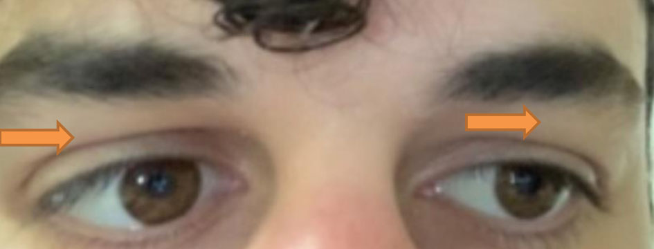 Click for large image | Figure 1. Eyelid edema in early mononucleosis known as Hoagland’s sign. Orange arrows indicate palpebral skin edema with evident flattening of the orbitopalpebral sulcus. |
He reported that about 15 days before the appearance of eyelid edema, he had nasal obstruction and purulent rhinorrhea, intense pharyngodynia and odynophagia accompanied by high fever (40 °C). The patient had taken antibiotic therapy in that circumstance. At first, he was treated with amoxicillin + clavulanic acid (orally, 1 g two times a day for 6 days), and subsequently with cefriaxone (intramuscular, 1 g a day for 6 days) with temporary remission of fever which reappeared a few days later with lower intensity (max. 38 °C).
Diagnosis
We subjected the patient to endoscopy of the upper airways and found clear external auditory canals, intact and rosy tympanic membranes, nasal septum in axis, hypertrophic inferior turbinates, free nasal foramina and recesses, free nasopharynx, normal oral cavity, and in oropharynx slight tonsillar hypertrophy with modest signs of acute inflammation. The glosso-epiglottic valleculae were free, and the piriform sinuses were free and distendable. The larynx was normal in morphology and motility. The cervical trachea was regular in appearance.
We then performed an ultrasound of the neck which showed the presence of multiple latero-cervical adenopathies of a reactive appearance with an elongated shape and preservation of the hilum. We found nothing significant in the thyroid and major salivary glands.
We diagnosed acute tonsillitis in progressive resolution associated with latero-cervical lymph node resentment. The endoscopic investigation allowed us to exclude acute rhinosinusitis as the cause of eyelid edema. The presence of sore throat and fever in the previous days and the presence at the time of the visit of modest signs of tonsillar inflammation, the finding of laterocervical adenopathies and the patient’s age, led us to suspect a form of infectious mononucleosis with possible Hoagland’s sign from disturbance of the lymphatic microcirculation and prescribed serological, blood chemistry (Table 1) and urine tests, also aimed at excluding any common water retention syndromes, possible causes of eyelid edema (glomerulonephritis, nephrotic syndromes, liver disease, thyroid disease).
 Click to view | Table 1. Results of the Required Blood Test |
The results of the tests requested confirmed the suspicion of infectious mononucleosis. We also requested an internal medicine evaluation during which abdominal ultrasound (which did not show hepatic or splenic involvement) and an infectious disease evaluation were also performed.
Treatment
Based on what emerged, we started oral therapy with prednisone (25 mg daily for 3 days and 12.5 mg for the next 3 days) and probiotic mixture [12, 13] for 1 month. The anti-inflammatory action of prednisone rapidly resolved the mild sore throat that still persisted at the time of our evaluation and contributed to the reduction of latero-cervical adenopathies. The patient no longer presented fever. We decided to resort to prednisone also for its anti-edema action in the hope of reducing eyelid edema. This however did not initially show a good response.
Follow-up and outcomes
The clinical picture progressively improved although eyelid edema persisted for 6 weeks.
| Discussion | ▴Top |
Infectious mononucleosis is the most frequent clinical expression of EBV infection. It is mainly characterized by a symptomatologic triad characterized by fever, sore throat, lymphadenopathy, lymphocytosis, and appearance of heterophile antibodies in serum [3].
In many cases, however, the infection can present an asymptomatic or paucisymptomatic course as well as begin or manifest during the course of the infection atypical symptoms.
Particularly interesting is the ocular involvement during the EBV infection with numerous possible clinical expressions. Librach in 1956 distinguished within a nucleus of patients affected by infectious mononucleosis two groups with different ocular manifestations [14]. The subjects with neuro-ophthalmic manifestations belonged to the first group, while the subjects with ocular adnexa manifestations belonged to the second group. In his work, the author claimed that conjunctivitis was the most frequent ocular manifestation. Since then, numerous authors have described ocular involvement during EBV infection [1, 2, 15]. Cases of hyperlacrimation, dacryoadenitis, dacryocystitis, hemorrhagic conjunctivitis, keratitis and still uveitis, choroiditis, retinitis, papillitis have been described.
In 1952, Hoagland was the first to describe the case of eyelid edema in mononucleosis. This sign was named after its first observer and became Hoagland’s sign [8]. He recognized this manifestation in a third of its patients with mononucleosis. However, this incidence does not appear realistic today as eyelid edema is a rare or even exceptional manifestation of the disease [16].
We performed a review of the cases of eyelid edema in mononucleosis reported in the literature since 1952. The scientific contributions were collected thanks to the PubMed search engine using as keywords: “Hoagland sign”, “ocular mononucleosis”, “eyelid edema mononucleosis”, “palpebral edema mononucleosis”, “dacryoadenitis mononucleosis”.
Table 2 shows the results of the research which confirm how this manifestation is very rare (28 cases described after the publication of the first case described by Hoagland in 1952) if we consider the very large diffusion of EBV infection in the population [17-30].
Click to view | Table 2. Cases of Hoagland’s Sign From 1952 to Present |
There are several hypotheses regarding the etiopathogenesis of this form of eyelid edema. The author agrees with Bass et al hypothesizing an impairment of lymphatic drainage as a possible causal mechanism of the manifestation and therefore supporting the association between eyelid edema and dacryoadenitis [1, 30]. According to other authors, the edema would be caused by a dacryocystitis due to the increase of B lymphocytes in the lymphatic tissues associated with the mucous membranes of the lacrimal glands [20].
Hoagland’s sign is usually temporary, and durations of a few days are predominantly described in the literature. In one case, the eyelid edema lasted for 8 weeks. In the case we described, the eyelid edema persisted for 6 weeks [30]. This makes it the second most long-lasting case described in the literature. Edema can be unilateral or bilateral, generally not painful, without skin hyperemia or visual changes.
The finding of eyelid edema in a patient poses the difficult issue of differential diagnosis. In the case described, we carried out blood tests and urinalysis which allowed us to rule out an edematous syndrome of renal or hepatic origin. We then investigated thyroid function to rule out a form of myxedema or other thyroid disease. In the case described, the occurrence of a syndrome characterized by pharyngodynia, high fever and latero-cervical lymph node reaction led to the immediate request for a blood count and a search for anti-EBV antibodies which resulted positive. Not having found results compatible with edema in water retention syndromes in the required blood chemistry tests, we definitively made the diagnosis of mononucleosis with dacryoadenitis and Hoagland’s sign. For this reason, we have avoided ascertaining the other possible causes of eyelid edema (ophthalmological, allergological, serological investigations, brain magnetic resonance imaging and orbits).
Conclusion
EBV is the etiological agent of mononucleosis, a syndrome frequently found in otorhinolaryngological clinics and usually characterized by some characteristic symptoms and signs: fever, general malaise, pharyngodynia accompanied by tonsillar hypertrophy, lymphocytosis and latero-cervical adenopathies. In some cases, mononucleosis can present a serious course if associated with some severe complications such as nephritis, myocarditis, hemolytic anemia, cranial nerve palsy and neuropathies, encephalitis and meningitis. In some rare cases of mononucleosis, impaired lymphatic drainage of the lacrimal glands can lead to the development of eyelid edema known as Hoagland’s sign. The recognition of this manifestation can favor an early diagnosis in the early stages of the disease.
Learning points
EBV is a widespread virus that causes frequent, in many cases asymptomatic, infections. Mononucleosis is the most frequent clinical syndrome encountered during EBV infection. Rarely, the disease can cause dacryoadenitis and consequent eyelid edema which poses various difficulties in terms of differential diagnostics but also possible delays in the recognition of the infection. The exceptional nature of this sign is confirmed by the literature review that we have carried out since 1952 (the date in which this sign was first described) and which revealed 28 cases before ours.
Acknowledgments
We acknowledge Dr.ssa Beatrice Bassi for support in English translation.
Financial Disclosure
No funding was received for this case report.
Conflict of Interest
The author declares no conflict of interest.
Informed Consent
The patient’s consent was requested and obtained for the processing of personal data and for the publication of the images.
Author Contributions
The manuscript was totally designed and written by the author MC.
Data Availability
The author declares that data supporting the findings of this study are available within the article.
Abbreviations
ALT: alanine aminotransferase; AST: aspartate aminotransferase; dL: deciliter; °C: degree Celsius; EBV: Epstein-Barr virus; ESR: erythrocyte sedimentation rate; FT4: thyroxine free; GGT: gamma glutamine transferase; Ig: immunoglobulins; L: liter; mg: milligram; mm: millimeter; µIU: international micro units; mL: milliliter; MRI: magnetic resonance imaging; PCR: C-reactive protein; pg: picogram; TSH: thyrotropin; U: unit; WBC: white blood cells
| References | ▴Top |
- Peponis VG, Chatziralli IP, Parikakis EA, Chaira N, Katzakis MC, Mitropoulos PG. Bilateral multifocal chorioretinitis and optic neuritis due to epstein-barr virus: a case report. Case Rep Ophthalmol. 2012;3(3):327-332.
doi pubmed pmc - Chervenkoff JV, Rajak SN, Brittain PG, Wright DA, Barrett VJ. Case report: a diagnostically challenging conjunctival mass caused by the Epstein-Barr virus. BMC Ophthalmol. 2015;15:129.
doi pubmed pmc - Womack J, Jimenez M. Common questions about infectious mononucleosis. Am Fam Physician. 2015;91(6):372-376.
pubmed - Luzuriaga K, Sullivan JL. Infectious mononucleosis. N Engl J Med. 2010;362(21):1993-2000.
doi pubmed - Grose CM. The many faces of infectious mononucleosis: the spectrum of Epstein-Barr virus infection in children. Pediatr Rev. 1985;7(2):35-44
- Wohl DL, Isaacson JE. Airway obstruction in children with infectious mononucleosis. Ear Nose Throat J. 1995;74(9):630-638.
pubmed - Putukian M, O'Connor FG, Stricker P, McGrew C, Hosey RG, Gordon SM, Kinderknecht J, et al. Mononucleosis and athletic participation: an evidence-based subject review. Clin J Sport Med. 2008;18(4):309-315.
doi pubmed - Hoagland RJ. Infectious mononucleosis. Prim Care. 1975;2(2):295-307.
pubmed - Hurt C, Tammaro D. Diagnostic evaluation of mononucleosis-like illnesses. Am J Med. 2007;120(10):911.e911-918.
doi pubmed - Bell AT, Fortune B, Sheeler R. Clinical inquiries. What test is the best for diagnosing infectious mononucleosis? J Fam Pract. 2006;55(9):799-802.
pubmed - Biggs TC, Hayes SM, Bird JH, Harries PG, Salib RJ. Use of the lymphocyte count as a diagnostic screen in adults with suspected Epstein-Barr virus infectious mononucleosis. Laryngoscope. 2013;123(10):2401-2404.
doi pubmed - Gelardi M, La Mantia I, Drago L, Meroni G, Aragona SE, Cupido G, Vicini C, et al. A probiotic mixture in patients with upper respiratory diseases: the point of view of the otorhinolaringologist. J Biol Regul Homeost Agents. 2020;34(6 Suppl 1):5-10.
pubmed - La Mantia I, Gelardi M, Drago L, Aragona SE, Cupido G, Vicini C, Berardi C, et al. Probiotics in the add-on treatment of pharyngotonsillitis: a clinical experience. J Biol Regul Homeost Agents. 2020;34(6 Suppl. 1):11-18.
pubmed - Librach IM. Ocular symptoms in glandular fever. Br J Ophthalmol. 1956;40(10):619-621.
doi pubmed pmc - Alba-Linero C, Rocha-de-Lossada C, Rachwani-Anil R, Sainz-de-la-Maza M, Sena-Corrales G, Romano V, Rodriguez-Calvo-de-Mora M. Anterior segment involvement in Epstein-Barr virus: a review. Acta Ophthalmol. 2022;100(5):e1052-e1060.
doi pubmed - Puntonieri E, Pastorino A, Di Bartolo CE, Leonardi V, Zagari D, Mileto G. Acute infectious mononucleosis presenting with palpebral and periorbital edema. Italian Journal of Medicine. 2012;6:227-230.
- Appelmans M, Van den Abeele L. [Acute dacryo-adenitis as a manifestation of infectious mononucleosis]. Bull Soc Belge Ophtalmol. 1967;146:249-258.
pubmed - Decker GR, Berberian BJ, Sulica VI. Periorbital and eyelid edema: the initial manifestation of acute infectious mononucleosis. Cutis. 1991;47(5):323-324.
pubmed - Marchese-Ragona R, Marioni G, Staffieri A, de Filippis C. Acute infectious mononucleosis presenting with dacryoadenitis and tonsillitis. Acta Ophthalmol Scand. 2002;80(3):345-346.
doi pubmed - Burger J, Thurau S, Haritoglou C. [Bilateral lid swelling during infectious mononucleosis (Hoagland-sign)]. Klin Monbl Augenheilkd. 2005;222(12):1014-1016.
doi pubmed - Inokuchi R, Iida H, Ohta F, Nakajima S, Yahagi N. Hoagland sign. Emerg Med J. 2014;31(7):561.
doi pubmed - Louppides S, Kakoullis L, Parpas G, Panos G. Upper eyelid oedema in a patient with pharyngitis/exudative tonsillitis and malaise: Hoagland sign in infectious mononucleosis. BMJ Case Rep. 2019;12(12):e233719.
doi pubmed pmc - Bonito FJP, Cerejeira D, Cunha H. Bilateral palpebral edema in a girl. Pediatr Dermatol. 2020;37(1):211-212.
doi pubmed - Kano Y, Kuki T. Young female patient with bilateral periorbital edema. Eur J Intern Med. 2020;75:93-94.
doi pubmed - Cherif MY, Richert B. Febrile palpebral edema. JAAD Case Rep. 2021;14:59-61.
doi pubmed pmc - Nakagawa H, Miyata Y, Maekawa M. Infectious mononucleosis with eyelid edema and palatal petechiae. Korean J Intern Med. 2021;36(4):1027-1028.
doi pubmed pmc - Otsuki T, Ishizuka K, Hirose M, Ie K. Hoagland sign in infectious mononucleosis. BMJ Case Rep. 2022;15(11):e252839.
doi pubmed pmc - Otsuka Y, Kishida M. Hoagland sign: bilateral upper eyelid oedema. BMJ Case Rep. 2022;15(6):e250857.
doi pubmed pmc - Bronz G, Zanetti B, Bianchetti MG, Milani GP, Lava SAG, Neuhaus TJ, Witschi A, et al. Bilateral upper eyelid swelling (Hoagland sign) in Epstein-Barr infectious mononucleosis: prospective experience. Infection. 2023;51(2):471-474.
doi pubmed pmc - Ricardo D. A protracted course of periorbital oedema in infectious mononucleosis caused by epstein-barr virus. Infect Dis Rep. 2022;14(6):942-945.
doi pubmed pmc
This article is distributed under the terms of the Creative Commons Attribution Non-Commercial 4.0 International License, which permits unrestricted non-commercial use, distribution, and reproduction in any medium, provided the original work is properly cited.
Journal of Medical Cases is published by Elmer Press Inc.