

| Journal of Medical Cases, ISSN 1923-4155 print, 1923-4163 online, Open Access |
| Article copyright, the authors; Journal compilation copyright, J Med Cases and Elmer Press Inc |
| Journal website http://www.journalmc.org |
Case Report
Volume 8, Number 4, April 2017, pages 105-107
Acute Pancreatitis: A Rare Presentation of Systemic Lupus Erythematosus
Tarana Guptaa, b, Ajit Singha, Rahul Chaudaa, Deepak Jaina, Hari K. Aggarwala
aDepartment of Medicine, Postgraduate Institute of Medical Sciences (PGIMS), Rohtak, Haryana, India
bCorresponding Author: Tarana Gupta, Department of Medicine, Postgraduate Institute of Medical Sciences (PGIMS), Rohtak, Haryana, India
Manuscript accepted for publication February 20, 2017
Short title: Acute Pancreatitis
doi: https://doi.org/10.14740/jmc2776w
| Abstract | ▴Top |
Systemic lupus erythematosus (SLE) is a systemic autoimmune disease with diverse clinical manifestations including gastrointestinal tract (GIT) afflictions. Lupus pancreatitis is more common in females and third decade of life with an incidence ranging from 0.7% to 4%. We here present a case of a 30-year-old female who presented with acute pancreatitis and after exclusion of all possible common etiology of pancreatitis, the evaluation revealed SLE with renal, skin and joint involvement. The patient showed marked improvement on immunosuppression with a resolution of all symptoms and proteinuria emphasizing the need for prompt diagnosis and management.
Keywords: SLE; Acute pancreatitis
| Introduction | ▴Top |
Systemic lupus erythematosus (SLE) is a systemic autoimmune inflammatory disease with diverse clinical manifestations. Gastrointestinal tract (GIT) afflictions may be due to the disease itself, adverse reactions of medications or by opportunistic infections. GIT involvement in SLE has various forms like mesenteric vasculitis (0.2-9.7%), protein-losing gastroenteropathy (1.9-3.2%), intestinal pseudo-obstruction which is rare, and lupus pancreatitis (0.7-4%) [1, 2]. Lupus pancreatitis is more common in females and third decade of life.
| Case Report | ▴Top |
A 30-year-old female resident of Punjab was admitted with complaints of arthralgia since 1 month and pain abdomen since 2 days. Arthralgias were intermittent, asymmetrical involving hands, wrists, and knees. She had epigastric pain which was sudden onset, rapidly progressive, radiating to back associated with abdominal distension and vomiting. The patient also had a history of photosensitivity and recurrent oral ulcers since 1 year.
On general physical examination, she had pallor and malar rash involving nasal bridge sparing the nasolabial folds. Her vitals were stable. There was no swelling, erythema, and deformity of joints. The abdomen was distended with the fullness of flanks and mild tenderness in the epigastric and periumbilical area and no appreciable organomegaly. Shifting dullness was present. Respiratory, cardiovascular and central nervous system examinations were unremarkable. Hematological investigations revealed microcytic, hypochromic anemia with hemoglobin of 8.7 g%. Kidney and liver function tests were essentially normal. Serum amylase (221 U/L; normal < 85 U/L) and lipase (349 U/L; normal < 60 U/L) were raised with a bulky and heterogenous echotexture of the pancreas with ascites on sonogram establishing a diagnosis of acute pancreatitis. She had no history of alcoholism, her sonography examination did not reveal any gall bladder stones and common bile duct was normal. Serum lipid profile, calcium and triglycerides levels were normal. Her urine examination revealed albuminuria with a 24-h protein of 2.5 g. Anti-nuclear antibodies (ANA) and anti-dsDNA antibodies were positive, with low C3 levels supporting a diagnosis of SLE, also serum proteins (5.4 g%; normal 6.5-8.5 g%) were decreased with an inverse albumin: globulin ratio (0.8). Ascitic fluid examination showed high protein (4.7 g%) and low serum-ascites albumin gradient (SAAG < 1.1) with raised amylase revealing pancreatic nature of ascites. Contrast CT scan of the abdomen was suggestive of acute pancreatitis with an acute pancreatic fluid collection of 4.5 × 2.1 cm size in the tail region of the pancreas and peripancreatic fat stranding (Fig. 1). Magnetic resonance cholangiopancreatography (MRCP) ruled out the possibility of congenital anomalies of the pancreas (Fig. 2). The patient improved with intravenous fluids and supportive management for pancreatitis. Later, her renal biopsy revealed evidence of mild mesangioproliferative glomerulonephritis suggestive of class II lupus nephritis. Subsequently, she was started on prednisolone 1 mg/kg/day dose with gradual tapering and ramipril 5 mg/day. Her ascites and abdominal pain improved with no recurrence and proteinuria decreased to < 500 mg/day.
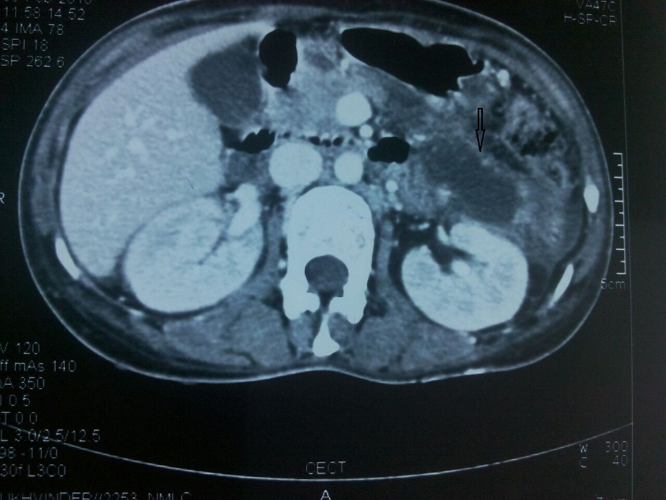 Click for large image | Figure 1. Axial contrast CT scan of the upper abdomen showing collection in relation to the tail of the pancreas. Thickening of left anterior pararenal, lateral conal and posterior pararenal fasciae is seen. |
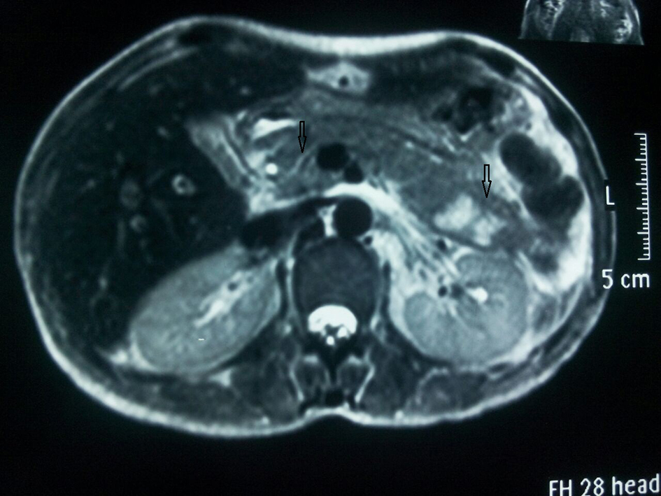 Click for large image | Figure 2. Collection in relation to the tail of the pancreas. The main pancreatic duct visualized in the head region has normal caliber and outline. |
| Discussion | ▴Top |
The incidence of acute pancreatitis in SLE varies from 0.7% to 4% and presents as initial manifestation in 22% and within first 2 years of diagnosis of SLE in 60% of all SLE-induced pancreatitis [1, 2]. Alcohol, gall stones, hypertriglyceridemia, hyperparathyroidism and congenital anomalies of pancreas like pancreas divisum and annular pancreas are common causes of pancreatitis which were ruled out in the index case. The pathophysiology of pancreatitis in SLE is multifactorial and may be related to complement activation and vasculitis with microthrombi formation in arteries and arterioles resulting in vascular damage or direct involvement of pancreatic parenchyma by autoantibodies and cellular immune response [1, 3]. The etiology may be acute pancreatitis as initial presentation or later during the course of disease due to either uncontrolled disease activity, drug toxicity or viral infection. The clinical manifestations of SLE pancreatitis can range from subclinical (an elevation of pancreatic enzymes without clinical symptoms) to acute pancreatitis or chronic pancreatitis (self-limiting) disease course. Patients with SLE and acute pancreatitis have higher systemic lupus erythematosus disease activity index (SLEDAI) score with increased incidence of multiorgan involvement like skin (46%), renal (35%), articular (43%), hematological (24%) and central nervous system (21%) [3, 4]. The index patient was a young female with pancreatitis as the first presentation of SLE with skin, articular and renal involvement. The lupus pancreatitis, unlike non-lupus pancreatitis, has higher incidence of leucopenia (59%) instead of leucocytosis [4]. Lupus pancreatitis is associated with high mortality up to 45% as compared to non-lupus pancreatitis with increased overall complications like recurrent pancreatitis (22%), respiratory failure (22%), pleural effusion (18%) and ascites (19%) [5]. The use of steroids has shown a reduction in mortality to 20% as compared to 61% when they are not used [4, 6]. Azathioprine although having a risk of developing pancreatitis has not been shown to increase the mortality in patients poorly controlled on steroids. We administered prednisolone in a dose of 1 mg/kg/day and tapered it to 10 mg/day and also started her on ramipril 5 mg/ day. The patient showed improvement in her symptoms with no recurrence of abdominal pain and decrease in proteinuria to < 500 mg/day.
Conclusion
SLE as a cause of pancreatitis should be actively considered in cases of pancreatitis where common etiologies have been excluded. Higher morbidity and mortality of SLE-induced pancreatitis and requirement of steroids and other forms of immunosuppression and disease modifying agents for management emphasize the need for high index of suspicion and early diagnosis. Steroids have shown to decrease mortality in this special population.
Author Contributions
TG, AL, RC, and HKA managed the patient. TG, AL, and DK wrote the manuscript. All authors approved the manuscript.
Financial Support
None.
Competing Interests
None.
Consent
Consent was obtained.
| References | ▴Top |
- Tian XP, Zhang X. Gastrointestinal involvement in systemic lupus erythematosus: insight into pathogenesis, diagnosis and treatment. World J Gastroenterol. 2010;16(24):2971-2977.
doi - Yuan S, Lian F, Chen D, Li H, Qiu Q, Zhan Z, Ye Y, et al. Clinical features and associated factors of abdominal pain in systemic lupus erythematosus. J Rheumatol. 2013;40(12):2015-2022.
doi pubmed - Pascual-Ramos V, Duarte-Rojo A, Villa AR, Hernandez-Cruz B, Alarcon-Segovia D, Alcocer-Varela J, Robles-Diaz G. Systemic lupus erythematosus as a cause and prognostic factor of acute pancreatitis. J Rheumatol. 2004;31(4):707-712.
pubmed - Nesher G, Breuer GS, Temprano K, Moore TL, Dahan D, Baer A, Alberton J, et al. Lupus-associated pancreatitis. Semin Arthritis Rheum. 2006;35(4):260-267.
doi pubmed - Wang CH, Yao TC, Huang YL, Ou LS, Yeh KW, Huang JL. Acute pancreatitis in pediatric and adult-onset systemic lupus erythematosus: a comparison and review of the literature. Lupus. 2011;20(5):443-452.
doi pubmed - Derk CT, DeHoratius RJ. Systemic lupus erythematosus and acute pancreatitis: a case series. Clin Rheumatol. 2004;23(2):147-151.
doi pubmed
This article is distributed under the terms of the Creative Commons Attribution Non-Commercial 4.0 International License, which permits unrestricted non-commercial use, distribution, and reproduction in any medium, provided the original work is properly cited.
Journal of Medical Cases is published by Elmer Press Inc.