

| Journal of Medical Cases, ISSN 1923-4155 print, 1923-4163 online, Open Access |
| Article copyright, the authors; Journal compilation copyright, J Med Cases and Elmer Press Inc |
| Journal website https://www.journalmc.org |
Case Report
Volume 14, Number 6, June 2023, pages 217-221

Chronic Unilateral Headache Related to Scar Neuromas
Karen S. Ferreiraa, c ![]() , Jose G. Specialib
, Jose G. Specialib
aDepartment of Neurology, Suroit Hospital, Salaberry-de-Valleyfield, QC, Canada
bDepartment of Neurology, Faculty of Medicine of Ribeirao Preto, University of Sao Paulo, Brazil
cCorresponding Author: Karen S. Ferreira, Department of Neurology, Suroit Hospital, Salaberry-de-Valleyfield, QC, Canada
Manuscript submitted April 3, 2023, accepted June 5, 2023, published online June 29, 2023
Short title: Chronic Unilateral Headache and Scar Neuromas
doi: https://doi.org/10.14740/jmc4087
| Abstract | ▴Top |
Postcraniotomy and posttraumatic headaches can result in scars generating local pain or referred pain following a neuropathic pattern. One hypothesis is that the pain can be caused and maintained by scar neuromas, developed after the nerve injury during the surgical process or trauma. This study reports two patients with chronic unilateral headaches: the first one with a posttraumatic scar in the parietal region and the other with a postsurgical scar in the mastoid region. In both patients, the headache was ipsilateral to the scar, suggesting primary headaches (trigeminal autonomic cephalalgia (TAC), as hemicrania continua and chronic cluster headache). Pharmacological treatment for these conditions failed. Instead, there was complete remission of the headache with anesthetic blockade of scar neuromas (demonstrated by clinical examination in both patients). An active search for traumatic or nontraumatic scars is recommended in all patients with refractory unilateral headaches, and anesthetic blocks for scar neuromas can be effective in treating this pain.
Keywords: Headache; Neuropathic pain; Nerve block; Chronic pain
| Introduction | ▴Top |
Surgical treatment or traumatic injury of any location in the body, can result in scars generating local pain or referred pain following a neuropathic pattern [1, 2].
This subtype of pain was not described by the International Classification of Diseases 10th Revision (ICD-10); but in the current version (International Classification of Diseases 11th Revision (ICD-11)), new chapters were added: chronic postsurgical pain (MG30.21) and chronic posttraumatic pain (MG30.20) [3, 4]. In both cases, the pain is localized in the surgical field or traumatic area, projected to the innervation territory or referred to a dermatome.
The best known model of postsurgical chronic pain is that related to the amputation of limbs (or part of them) and postsurgical low back chronic pain, also called “failed back surgery syndrome (FBSS)”, located in the surgical site or projected to the limbs [4]. About 10% to 40% (average 20%) of all patients undergoing low back surgery develop some form of chronic pain that requires additional low back surgery, pain-related medical consultations, or other surgical interventions such as neuromodulation to deal with pain [1]. Chronic low back pain after surgery is reported as severe by 13% of patients after spinal stenosis and herniated disc surgery. Other types of postsurgical pain include thoracotomies, mastectomies, hysterectomies, hip and knee surgeries and amputations [4], followed by head and neck surgeries (more than 50% of these injuries are related to tooth extraction [2].
Chronic postsurgical pain can be caused and maintained by scar neuromas, developed 1 to 12 months after cutting the nerves during the surgical process [5, 6]. On the other hand, chronic posttraumatic pain can also arise after nerve damage due to pressure, crush injuries, cuts, lacerations, stretching, bleeding into the tissue surrounding the nerve, irritation produced by foreign material in contact with a nerve, infections, ischemia , by inclusion of nerves in the ligation of an artery, fracture of the bone canal, nerve fibers pressed by nearby structures and in fractures when the fragments are misaligned [7]. The pain, therefore, would be generated not only by surgical amputation neuromas [7]. Therefore, it is possible that some headaches as cervicogenic headaches, occipital neuralgia [8], classic trigeminal neuralgias, auriculotemporal neuralgias, supraorbital neuralgias, carpal tunnel syndrome, among others, may be other examples of posttraumatic chronic pain generated by mechanisms above. This would explain why chronic pain caused by surgery, trauma, and other clinical conditions improves with local blocks.
This study reports two patients with chronic unilateral headaches: the first one with a posttraumatic scar in the parietal region and the other with a postsurgical scar in the mastoid region. In both patients, the headache was ipsilateral to the scar, and it was resolved after an anesthetic block.
| Case Reports | ▴Top |
Case 1
Investigations
A 34-year-old male was referred by the primary health service for treatment at the Headache Clinic, due to left hemicrania headache for 2 years, with no improvement in the pain with treatments performed. The headache started with low intensity and increased over months. Currently, it was a pressure pain, unilateral, and continuous. In addition of this continuous headache, he described paroxysmal episodes of pain, three to four times a day, stabbing, prickling or throbbing, lasting 1 hour, with sense of restlessness and agitation during the pain, associated with mild nausea, without photophobia or phonophobia. Brain magnetic resonance imaging (MRI) was normal.
Diagnosis
Posttraumatic headache mimicking hemicrania continua, but there was no response to indomethacin test.
Treatment
The indomethacin was maintained for 2 months at doses between 100 to 200 mg per day, with no response. After that, several therapeutic approaches, pharmacological (e.g., topiramate, amitriptyline) and non-pharmacological, were performed without any clinical improvement, during 2 years of follow-up. On a given day, the patient appeared with shaved hair (he had thick and long hair before) and there was a huge scar from a previous head trauma, which had occurred about 8 months before the onset of the headache. The scar had not been reported in previous consultations. Examining the scar, it was observed small nodules measuring 1 - 2 mm, which submitted to digital pressure, triggered the same lancinating, stabbing pain, which was followed, after minutes, by an increased pain (Fig. 1, nodules marked). An anesthetic block was performed, injecting approximately 0.1 - 0.2 mL of dexamethasone 10% and 0.4 mL of lidocaine 2% without vasoconstrictor, in each diagnosed nodule.
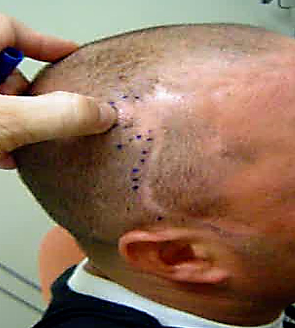 Click for large image | Figure 1. Scar from a previous head trauma (case 1). |
Follow-up and outcomes
The pain improved, with no relapse, until the return, a month later. The pain triggers had disappeared and finally the patient left the Headache Clinic.
Case 2
Investigations
A 37-year-old male was referred by the primary health service for treatment at the Headache Clinic, with a chronic severe headache, right-sided, with autonomic signs. The patient reported that the headache started 2 years before. It was completely resolved in 1 - 2 months, disappeared for more than a year and then it relapsed. The headache was right-sided, severe, excruciating and usually woke him up in the middle of the night. It was completely improved in 1 - 2 h, but it could be repeated one to three times in the same day, also occurring during wakefulness with the same characteristics and at the same time. He reported that during the attack he was very agitated, with red eye, tearing and, sometimes, a runny nose. The pain has been persisting daily throughout this time. Clinical examination showed a right eye miosis and an important surgical scar from a right-sided mastoidectomy in the past. Some small nodules measuring 1 - 2 mm, and very painful to digital pressure, were present on the edges of the surgical scar. (Fig. 2). Brain MRI was performed, which was normal.
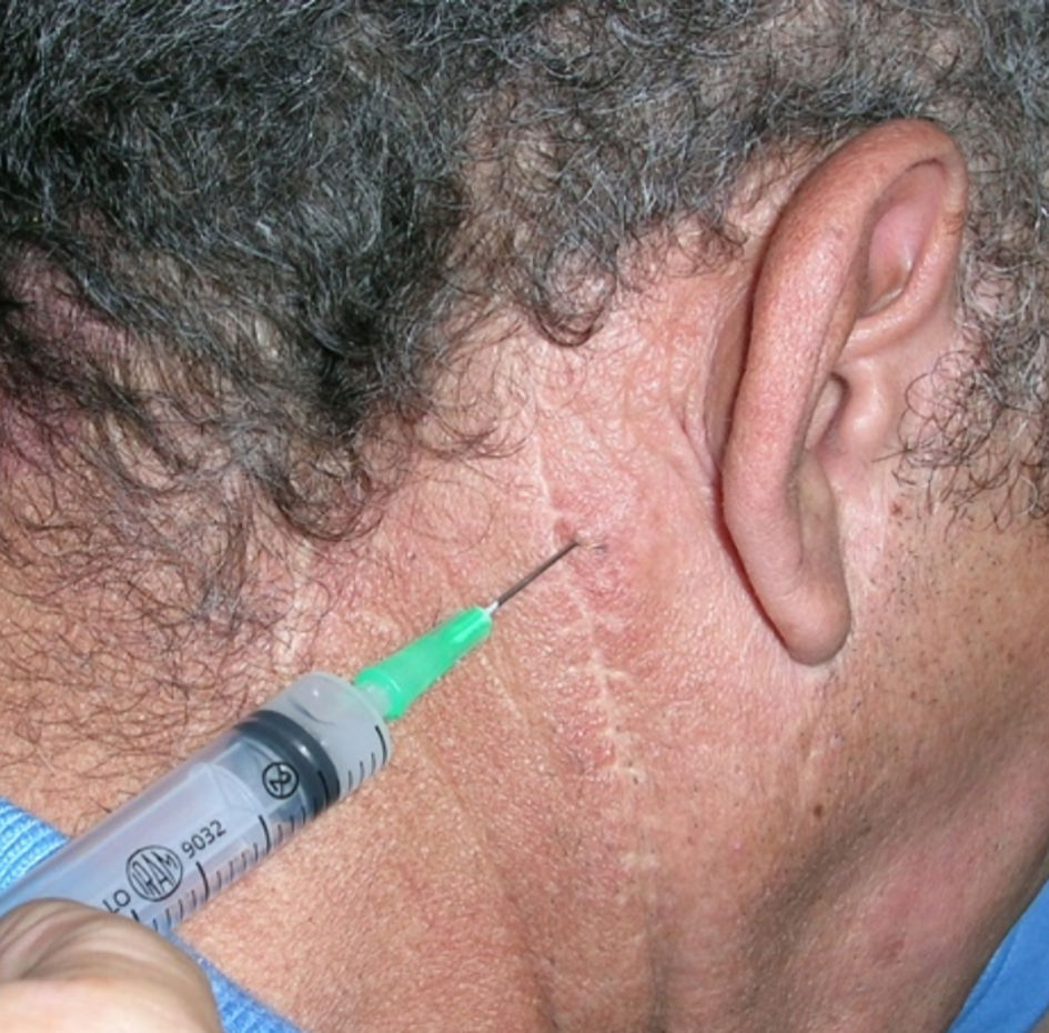 Click for large image | Figure 2. Scar from a right-sided mastoidectomy (case 2). |
Diagnosis
The diagnosis was postcraniotomy headache with features of chronic cluster headache.
Treatment
Thus, treatment for cluster headache was instituted, including verapamil 320 mg/day (increased to 480 mg/day) and oral corticoid. There was no improvement with this treatment, nor with the combination of topiramate (150 mg/day) and melatonin (10 mg at night). It was then decided to block the small nodules in the surgical scar with an injection of approximately 0.1 - 0.2 mL of dexamethasone 10% and 0.4 mL of lidocaine 2% without vasoconstrictor in each trigger point.
Follow-up and outcomes
It improved until the next follow-up (1 month), allowing the withdrawal of the medications gradually until the interruption. Three months later he returned without headache.
| Discussion | ▴Top |
Herein two case reports were described with clinical conditions that suggested diagnostic criteria for primary headache (trigeminal autonomic cephalalgia (TAC)), one of them suggesting hemicrania continua (but without indomethacin response) and the other suggesting chronic cluster headache. Nevertheless, pharmacological treatment for these conditions failed. Instead, there was complete remission of the headache with anesthetic blockade of scar nodules (demonstrated by clinical examination in both patients), one with a posttraumatic scar and the other with a postsurgical scar.
We believe that both patients described in this study presented neuromas in skull scars. Neuromas can be diagnosed in scars by digital pressure (nodules of 1 - 2 mm at the edges of the scars) that trigger intense stinging pain and/or the presence of referred pain (burning, stabbing or continuous) in the areas corresponding to the peripheral distributions of the nerves. On examination, changes in local sensitivity (hypoesthesia, hyperalgesia or anesthesia) can also be verified [8]. Neuromas usually present as a firm, oval, slowly growing nodule no larger than 2 cm. A prominent area visible in the scar may be a characteristic of a neuroma [7, 8].
In this study, patients did not report pain in the scar itself. They reported headaches with autonomic signs, one with hemicrania continua phenotype and the other with cluster headache phenotype. This situation could be understood considering the possibility of central sensitization [9, 10]. Nociceptive afferents originated in the scar, which can be clinical or subclinical (in these two cases subclinical), reaching the brain, in specific sites, would demodulate the mechanisms related to the pathophysiology of trigeminal autonomic headaches, triggering symptoms phenotypically like the primary headaches; but in this case, they were secondary headaches. Once the nociceptive afferents in the scar neuromas were blocked, the headaches disappeared.
According to the International Classification of Headache Disorders (ICHD) [11], headache related to traumatic scar (first patient) would be classified as “5.2 persistent headache attributed to traumatic head injury”, but it did not meet all diagnostic criteria. It can be better classified as “Appendix (A5.2) persistent headache attributed to traumatic head injury”. The difference between the two proposals, one found in the body of the classification and the other in the appendix, is the time for the onset of chronic persistent headache. Appendix (A5.2) admits that this time may be longer than 3 months. Interestingly, the initial diagnosis was possibly hemicrania continua, but finally there was no response to indomethacin test.
Furthermore, according to ICHD [11], the headache related to the surgical scar (second patient) would be classified as “5.6 persistent headache attributed to craniotomy”, without fulfilling all diagnostic criteria. Otherwise, this headache phenotype met criteria for chronic cluster headache (“3.1.2 chronic cluster headache”). Literature review was performed, and it did not report posttraumatic or postcraniotomy headache with the phenotype of TAC.
Regarding treatment, since pharmacological treatment did not improve the symptoms, the option to treat our patients was the anesthetic block of the scar neuromas, observed by physical examination. All diagnosed neuromas were blocked. The response to anesthetic block was described before and it can explain the decision for this conduct [12-14].
Previous literature described pharmacological treatment for neuromas, including N-methyl-D-aspartate (NMDA) receptor antagonists, opioids, anticonvulsants, antidepressants, local anesthetics, and calcitonin, which act by inhibiting pain signaling pathways and central and peripheral sensitization. For patients who develop pain that is refractory to pharmacological treatment, alternative intervention is described, such as cryotherapy and spinal cord stimulation, but they are ineffective and may cause undesirable consequences.
Finally, for patients with intractable painful neuromas, surgery may be the best choice. In general, capping the nerve end or covering it with muscle or bone flaps is strongly recommended for all patients with painful neuromas, as this approach not only reduces mechanical stimulation, but also isolates the skin and scar tissue from the neuroma, thus avoiding pain. Another effective treatment includes ligation and restoration of nerve continuity. These cases could be treated surgically from the beginning and the period of patient suffering can be shortened.
On the other hand, postcraniotomy headache can also be treated with sub-anesthetic ketamine infusion or surgical site injection with local anesthetics, corticosteroids, or botulinum toxin [15, 16].
The treatment proposed in this study was the anesthetic injection of all neuromas identified in the scars, due to our team experience with this treatment [14]. Although numerous treatments have been proposed for painful neuromas, including prevention techniques, non-surgical treatment, and surgical treatment, none of them are universally accepted and they usually provide incomplete pain relief, inducing side effects. This reflects the frustrating clinical prognosis and high failure rates of all these treatment methods [13].
A previous study was published by our team, describing postsurgical unilateral headaches [14]. Four patients were described (one had a benign brain tumor, two had intracranial aneurysm surgery and one had temporal lobectomy for epilepsy). All of them presented hemicrania after a variable time interval (from 1 month to 9 years). Migraine-like and trigeminal-autonomic symptoms (without criteria for TAC) related to scar neuromas were described.
The limitation of this study was the short follow-up time of the patients. As our university hospital is considered by the population to be the last hope for unresolved problems elsewhere, if patients did not return for a long time, after neuroma treatment for follow-up visits, our conclusion is that the treatment was successful. However, a long-term follow-up would be suitable for these patients.
Conclusions
Chronic posttraumatic and postcraniotomy headache treatment can be a challenge in clinical practice. One hypothesis is that the pain can be caused and maintained by scar neuromas. Otherwise, an active search for traumatic or nontraumatic scars is recommended in all patients with refractory unilateral headaches, and anesthetic blocks for scar neuromas can be effective in treating this pain.
Learning points
The most important conclusions related to this study can be summarized here: 1) Persistent unilateral headaches, with specific or unspecific characteristics, may be secondary to trauma or surgery; 2) An active search for traumatic or nontraumatic scars (from herpes zoster, for example) is recommended in all patients with refractory unilateral headaches; 3) The time interval between the trauma and the onset of chronic pain may be longer than accepted in ICHD; 4) Anesthetic blocks for scar neuromas can be effective in treating posttraumatic and postsurgical headaches.
Acknowledgments
None to declare.
Financial Disclosure
None to declare.
Conflict of Interest
None to declare.
Informed Consent
Informed consent was obtained.
Author Contributions
Karen S. Ferreira: discussion, writing the paper, and final approval. Jose G. Speciali: design, data collection, discussion, writing the paper, and final approval.
Data Availability
The authors declare that data supporting the findings of this study are available within the article.
| References | ▴Top |
- Yao C, Zhou X, Zhao B, Sun C, Poonit K, Yan H. Treatments of traumatic neuropathic pain: a systematic review. Oncotarget. 2017;8(34):57670-57679.
doi pubmed pmc - Thomas DC, Mallareddy SD, Okeson JP, Thankachan J, Pitchumani PK, Pichammal RC. Trigeminal traumatic neuroma: a comprehensive review of the literature based on a rare case. Curr Pain Headache Rep. 2022;26(3):219-233.
doi pubmed - Korwisi B, Treede RD, Rief W, Barke A. Evaluation of the International Classification of Diseases-11 chronic pain classification: study protocol for an ecological implementation field study in low-, middle-, and high-income countries. Pain Rep. 2020;5(4):e825.
doi pubmed pmc - Schug SA, Lavand'homme P, Barke A, Korwisi B, Rief W, Treede RD, Pain ITftCoC. The IASP classification of chronic pain for ICD-11: chronic postsurgical or posttraumatic pain. Pain. 2019;160(1):45-52.
doi pubmed - Murphey MD, Smith WS, Smith SE, Kransdorf MJ, Temple HT. From the archives of the AFIP. Imaging of musculoskeletal neurogenic tumors: radiologic-pathologic correlation. Radiographics. 1999;19(5):1253-1280.
doi pubmed - Oliveira KMC, Pindur L, Han Z, Bhavsar MB, Barker JH, Leppik L. Time course of traumatic neuroma development. PLoS One. 2018;13(7):e0200548.
doi pubmed pmc - Rajput K, Reddy S, Shankar H. Painful neuromas. Clin J Pain. 2012;28(7):639-645.
doi pubmed - Choi I, Jeon SR. Neuralgias of the head: occipital neuralgia. J Korean Med Sci. 2016;31(4):479-488.
doi pubmed pmc - Chau MN, Jonsson E, Lee KM. Traumatic neuroma following sagittal mandibular osteotomy. Int J Oral Maxillofac Surg. 1989;18(2):95-98.
doi pubmed - Arendt-Nielsen L, Morlion B, Perrot S, et al. Assessment and manifestation of central sensitization across different chronic pain conditions. Eur J Pain. 2018;22:216-241.
- The international classification of headache disorders, 3rd edition. Cephalalgia. 2018; 38:1-211.
- Lovely TJ. The treatment of chronic incisional pain and headache after retromastoid craniectomy. Surg Neurol Int. 2012;3:92.
doi pubmed pmc - Lu C, Sun X, Wang C, Wang Y, Peng J. Mechanisms and treatment of painful neuromas. Rev Neurosci. 2018;29(5):557-566.
doi pubmed - Ferreira Kdos S, Dach F, Speciali JG. Scar neuromas as triggers for headache after craniotomy: clinical evidence. Arq Neuropsiquiatr. 2012;70(3):206-209.
doi pubmed - Lutman B, Bloom J, Nussenblatt B, Romo V. A contemporary perspective on the management of post-craniotomy headache and pain. Curr Pain Headache Rep. 2018;22(10):69.
doi pubmed - Chappell AG, Yuksel S, Sasson DC, Wescott AB, Connor LM, Ellis MF. Post-mastectomy pain syndrome: an up-to-date review of treatment outcomes. JPRAS Open. 2021;30:97-109.
doi pubmed pmc
This article is distributed under the terms of the Creative Commons Attribution Non-Commercial 4.0 International License, which permits unrestricted non-commercial use, distribution, and reproduction in any medium, provided the original work is properly cited.
Journal of Medical Cases is published by Elmer Press Inc.