

| Journal of Medical Cases, ISSN 1923-4155 print, 1923-4163 online, Open Access |
| Article copyright, the authors; Journal compilation copyright, J Med Cases and Elmer Press Inc |
| Journal website http://www.journalmc.org |
Case Report
Volume 9, Number 6, June 2018, pages 160-163
A Rare Presentation of Large Cell Neuroendocrine Carcinoma of the Lung
Ryan Dixona, b, Nada Mohameda, Thomas Genesea
aUnited Health Services Hospitals, Inc, Johnson City, NY, USA
bCorresponding Author: Ryan Dixon, United Health Services Hospitals, Inc., Johnson City, NY 13790, USA
Manuscript submitted March 22, 2018, accepted April 3, 2018
Short title: Large Cell Neuroendocrine Carcinoma of the Lung
doi: https://doi.org/10.14740/jmc3047w
| Abstract | ▴Top |
Large cell neuroendocrine carcinoma of the lung is a rare form of pulmonary tumor, representing 2-3% of all non-small cell carcinomas. The large majority of patients who are diagnosed with this disease have a smoking history. We present a non-smoking patient with no prior medical history who presented with recurrent stomach pain and scalp lesions who was ultimately diagnosed with large cell neuroendocrine carcinoma of the lung. Due to its rarity, there is little consensus on effective treatment and despite progressive therapy with currently recommended regimens, the patient ultimately passed from complications of the disease. The fact that the patient developed a form of lung cancer so highly associated with smoking history coupled with the uncommon presentation in such advanced disease is exceedingly rare.
Keywords: Large cell neuroendocrine carcinoma of the lung; Large cell neuroendocrine carcinoma; Neuroendocrine carcinoma; Pulmonary tumor; Non-small cell carcinomas
| Introduction | ▴Top |
Lung cancer is the leading cause of cancer death in both men and women in the United States. Approximately 90 percent of all lung cancers are associated with cigarette smoking. While lung cancer can be divided into several histologic subtypes, the most important distinction is between small cell lung cancer and non-small cell lung cancer. Large cell neuroendocrine carcinoma (L-LCNEC) of the lung is a rare form of pulmonary tumor, representing 2-3% of all non-small cell carcinomas. The large majority of patients who are diagnosed with this disease have a smoking history. At the time of diagnosis, lung cancer is typically in an advanced stage. Presenting symptoms typically include cough, hemoptysis, dyspnea, and chest pain. Common sights of metastases are brain, bone, and liver, and adrenal glands.
We present the rare case of a patient with no previous symptoms typical of pulmonary tumor who presented with recurrent stomach pain and lesions of the scalp. Subsequent biopsies of a suspicious gastric ulcer and the scalp lesions revealed the patient had advanced L-LCNEC. Despite treatment with currently recommended regimens, the patient ultimately passed from complications of the disease.
| Case Report | ▴Top |
This is a previously healthy 47-year-old male, never smoker, who presented to his primary care provider with 2 months of upper abdominal pain that was dull, moderate in severity, usually occurring in the morning, and was eased by eating. He denied other symptoms including weight loss, fevers, chills, sweats, blood in stool, nausea, vomiting, constipation, diarrhea, and change in appetite. Other than positive fecal occult blood test the physical exam was within normal limits. At that time he was started on oral omeprazole 20 mg daily and his symptoms improved temporarily. Two months later, he presented to his primary care provider again with recurrent abdominal pain and a new complaint of 6 lb weight loss since the last office visit. Omeprazole was increased to 40 mg oral daily and he was referred to gastroenterology.
Two weeks later (and before the gastroenterology referral) he visited a walk-in clinic because he noticed four lesions on his scalp. He was given a methylprednisolone dose pack, which he did not take, and referred to dermatology. He was seen 2 weeks later and the dermatologist described the lesions as inflammatory papules of unknown cause, exquisitely tender with the oldest lesion being 6.0 mm in diameter and 2.0 mm high. Other nodules were between 4.0 and 5.0 mm in diameter. Incision and drainage of the largest nodule was attempted and yielded only small drops of blood. The dermatologist noted that the pain and rapid onset were unusual. Differential diagnosis included granulomas, scars, and adnexal tumors. The patient was scheduled for a biopsy.
Two days after being seen by a dermatologist, the patient underwent upper endoscopy for his abdominal pain. It was noted that the esophagus and gastroesophageal junction appeared normal. Upon advancing into the stomach the gastric mucosa was found to be diffusely erythematous. There was a gastric ulcer with elevated borders in the mid gastric body along the posterior wall that was suspicious for malignancy (Fig. 1). Biopsies of the lesion were taken and the rest of the endoscopy was found to be unremarkable. He was instructed to continue oral omeprazole 40 mg daily and advised to avoid non-steroidal anti-inflammatory drugs (NSAIDs). The following week the patient underwent incisional biopsy of the scalp lesions under local anesthesia.
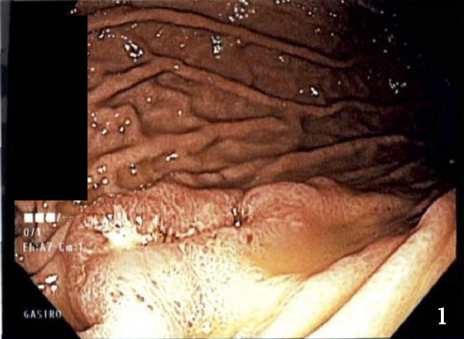 Click for large image | Figure 1. Photograph of stomach ulcer taken during upper endoscopy. |
The biopsies of the stomach ulcer and scalp lesions showed a neuroendocrine tumor. Both sites stained positively for cytokeratin 7 (CK7), synaptophysin, chromogranin, CD56, thyroid transcription factor-1 (TTF-1) and weak caudal-related homeobox transcription factor 2 (CDX-2). Stomach biopsy showed six mitoses/10 HPF and Ki-67 stain showed proliferation rate of approximately 40%. Scalp biopsy showed 11 mitoses/ 10 HPF and a proliferation rate of 55%. The biopsy results were sent to the University of Rochester Medical Center, Strong Memorial Hospital for a second opinion at the patient’s request; the results were confirmed. These findings suggested a possible lung primary with metastases to the stomach and scalp. The patient underwent a positron emission tomography (PET) scan, which revealed a 2.8 cm lung mass with hypermetabolic left hilar adenopathy suggestive of a primary lung neoplasm. It also showed multiple tumors in the liver and bones.
The patient subsequently underwent treatment with etoposide and cisplatin. Despite chemotherapy his disease continued to progress. Follow-up CT scans of the chest, abdomen, and pelvis revealed progression of the disease. The scalp lesions continued to increase in size and associated pain so he was referred to Radiation Oncology for possible radiation. Due to the number of lesions it was determined that whole brain radiation therapy was indicated, and the patient underwent 10 treatments. Due to failure of the primary chemotherapy regimen the patient was placed on an alternative regimen with capecitabine with temozolomide. Upon failure of that regimen, he was placed on crizotinib since he had the echinoderm microtubule-associated protein-like 4-anaplastic lymphoma kinase (EML4-ALK) translocation. Upon failure of that medication, he was also put on a kinase inhibitor. Despite multiple regimens the disease continued to progress, and approximately 2 years after diagnosis the patient passed away due to complications of the disease.
| Discussion | ▴Top |
Large-cell neuroendocrine carcinoma of the lung is a rare disease, making up just 2-3% of all non-small cell carcinoma [1]. L-LCNEC is typically diagnosed at earlier stages than other forms of lung cancer and usually located peripherally. Diagnosis is based on the 2015 World Health Organization (WHO) classification which includes neuroendocrine morphology, high mitotic rate (> 10/2 mm2 with median of 70/2 mm2), necrosis, cytologic features (e.g., large cell size, low nuclear to cytoplasmic ratio, vesicular or fine chromatin, or frequent nucleoli), and positive immunohistochemical staining for one or more neuroendocrine markers or granules by electron microscopy. Typical cellular neuroendocrine morphological features include organoid, nesting palisading rosettes, and trabeculae (Fig. 2, 3). Mitotic rates are usually > 10/10 HPF which was demonstrated in the biopsy of the scalp lesions. Cells are large in size, have low nuclear to cytoplasmic ratio, fine chromatin, and frequent nucleoli. Finally, immunohistochemical staining is positive for one or more neuroendocrine markers, usually CD56, synaptophysin, and chromogranin, all of which were demonstrated on staining of both the stomach ulcer and scalp lesions (Fig. 4, 5) in our patient.
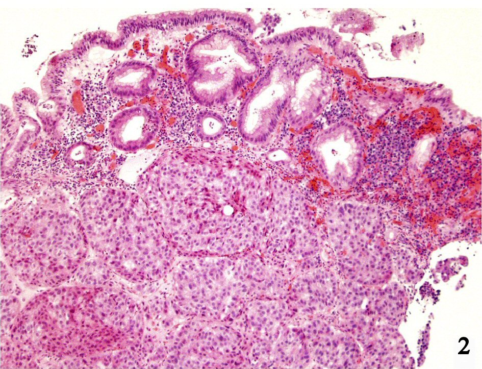 Click for large image | Figure 2. Gastric mucosa with underlying nests of large cells (H&E; × 100). |
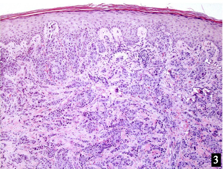 Click for large image | Figure 3. Skin with dermal nests of large cells (H&E; × 100). |
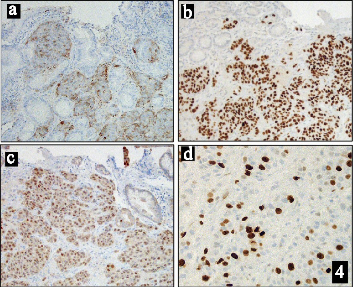 Click for large image | Figure 4. Immunohistochemical stains of: (a) Chromogranin (× 100). (b) TTF1 (× 100). (c) CDX2 (× 100). (d) Ki67 (× 200). |
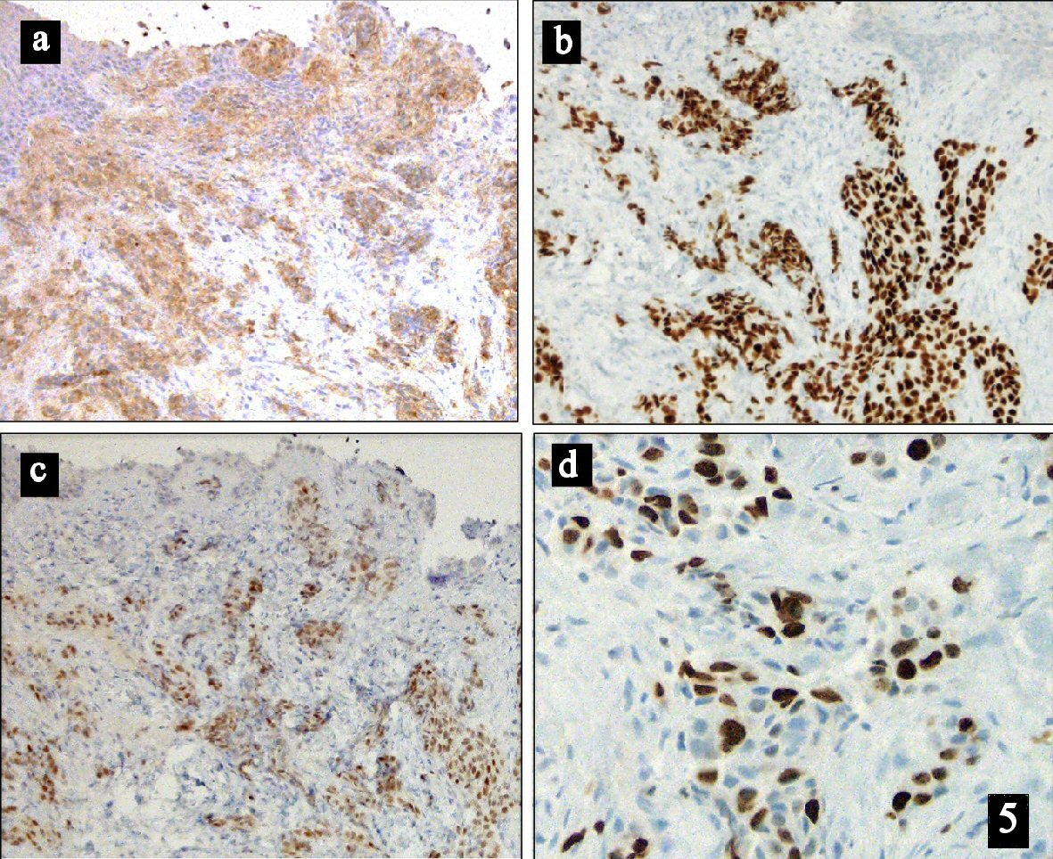 Click for large image | Figure 5. Immunohistochemical stains of: (a) Chromogranin (× 100). (b) TTF1 (× 100). (c) CDX2 (× 100). (d) Ki67 (× 200). |
The most significant risk factor for developing lung cancer, including large cell neuroendocrine carcinoma, is smoking [2]. One retrospective study showed that 98.6% of patients with diagnosis of large-cell neuroendocrine carcinoma of the lung had extensive past or present smoking histories [3]. The fact that the patient developed a form of lung cancer so highly associated with smoking history coupled with the uncommon presentation in such advanced disease makes this patient unique.
Due to its rarity, there is little consensus on effective treatment [1]. Currently treatment is guided by staging, with early stages (I-II) are typically surgically resected with mediastinal lymph node sampling or dissection. Later stages (III-IV) treated similarly to small cell carcinoma of the lung with etoposide and cisplatin [4]. However, even with these treatment regimens, prognosis remains poor. This patient did not respond well to first line therapy, nor did he respond to subsequent therapies.
This case demonstrates a rare presentation of lung cancer. While the disease was very advanced at diagnosis, the patient manifested symptoms and signs that are atypical of lung cancer, namely abdominal pain and scalp lesions, which are uncommon areas of metastases.
| References | ▴Top |
- Glisson BS, Moran CA. Large-cell neuroendocrine carcinoma: controversies in diagnosis and treatment. J Natl Compr Canc Netw. 2011;9(10):1122-1129.
doi pubmed - Alberg AJ, Samet JM. Epidemiology of lung cancer. Chest. 2003;123(1 Suppl):21S-49S.
doi pubmed - Asamura H, Kameya T, Matsuno Y, Noguchi M, Tada H, Ishikawa Y, Yokose T, et al. Neuroendocrine neoplasms of the lung: a prognostic spectrum. J Clin Oncol. 2006;24(1):70-76.
doi pubmed - Le Treut J, Sault MC, Lena H, Souquet PJ, Vergnenegre A, Le Caer H, Berard H, et al. Multicentre phase II study of cisplatin-etoposide chemotherapy for advanced large-cell neuroendocrine lung carcinoma: the GFPC 0302 study. Ann Oncol. 2013;24(6):1548-1552.
doi pubmed
This article is distributed under the terms of the Creative Commons Attribution Non-Commercial 4.0 International License, which permits unrestricted non-commercial use, distribution, and reproduction in any medium, provided the original work is properly cited.
Journal of Medical Cases is published by Elmer Press Inc.