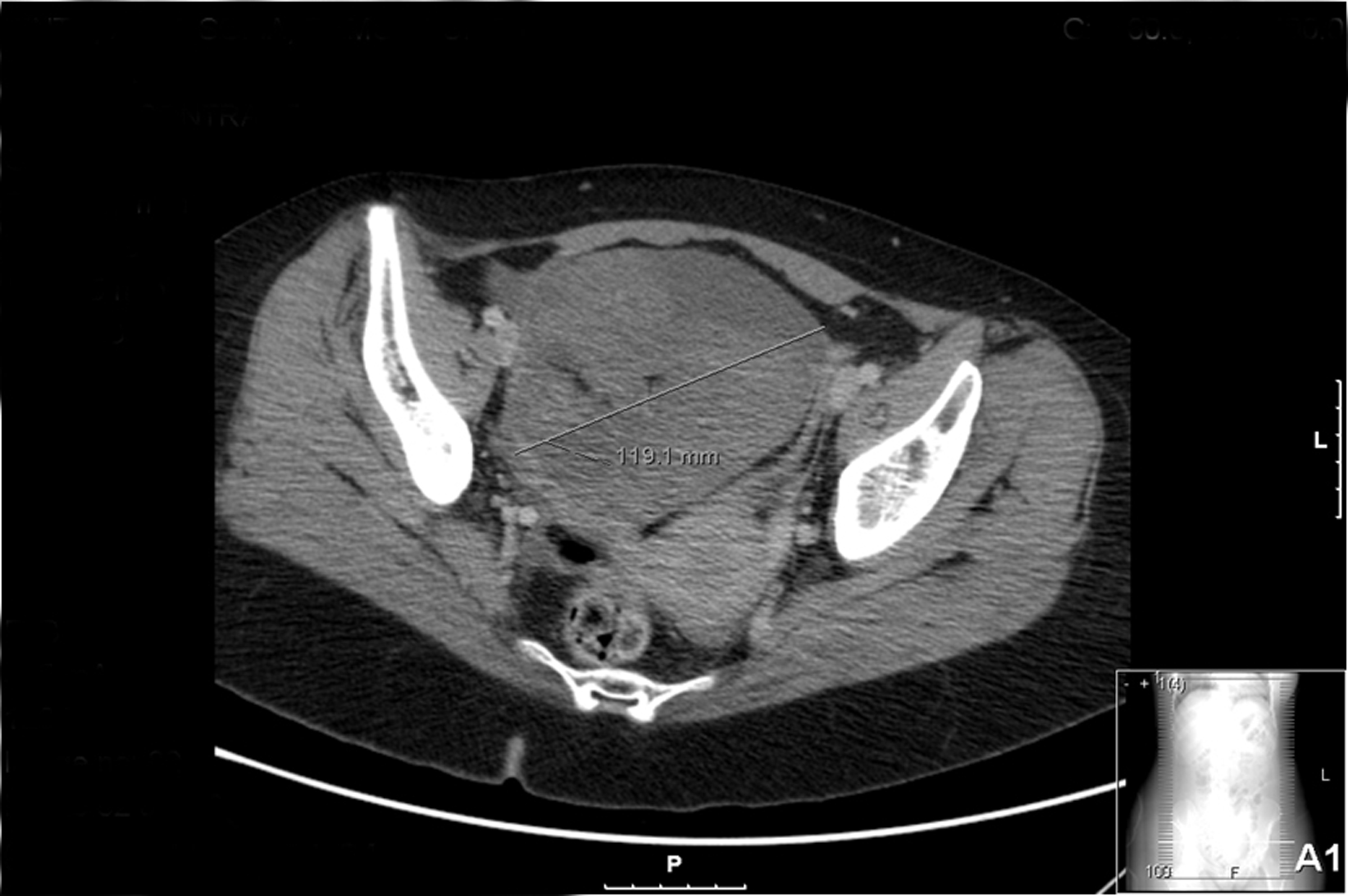
Figure 1. CT scan of the patient revealed a pelvic mass located to the right ovary with 119 × 132 mm in dimensions; calcification and fat foci inside suggestive of malignancy namely teratoma.
| Journal of Medical Cases, ISSN 1923-4155 print, 1923-4163 online, Open Access |
| Article copyright, the authors; Journal compilation copyright, J Med Cases and Elmer Press Inc |
| Journal website http://www.journalmc.org |
Case Report
Volume 4, Number 3, March 2013, pages 135-138
Anti-NMDA Receptor Paraneoplastic Encephalitis: An Important Differential Diagnosis in Subacute Psychosis
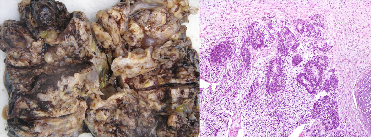
Figures