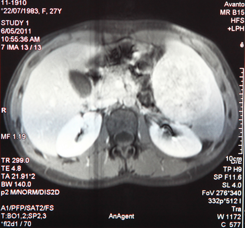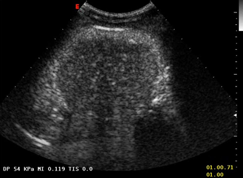
Figure 1. Hypohecoic rounded lesion appearing after echo amplifier (sulphur hexafluoride) injection.
| Journal of Medical Cases, ISSN 1923-4155 print, 1923-4163 online, Open Access |
| Article copyright, the authors; Journal compilation copyright, J Med Cases and Elmer Press Inc |
| Journal website http://www.journalmc.org |
Case Report
Volume 4, Number 2, February 2013, pages 75-79
Inflammatory Pseudotumor of the Spleen: A Case Report and Literature Review
Figures