Journal of Medical Cases, ISSN 1923-4155 print, 1923-4163 online, Open Access
Article copyright, the authors; Journal compilation copyright, J Med Cases and Elmer Press Inc
Journal website http://www.journalmc.org

Case Report

Volume 4, Number 3, March 2013, pages 146-148


Proptosis, Diplopia, and an Ocular Bruit in a Patient With Ehlers-Danlos Syndrome

Figures

Figure 1.
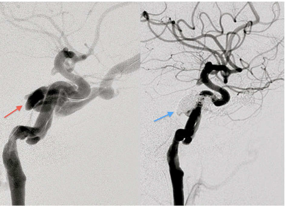
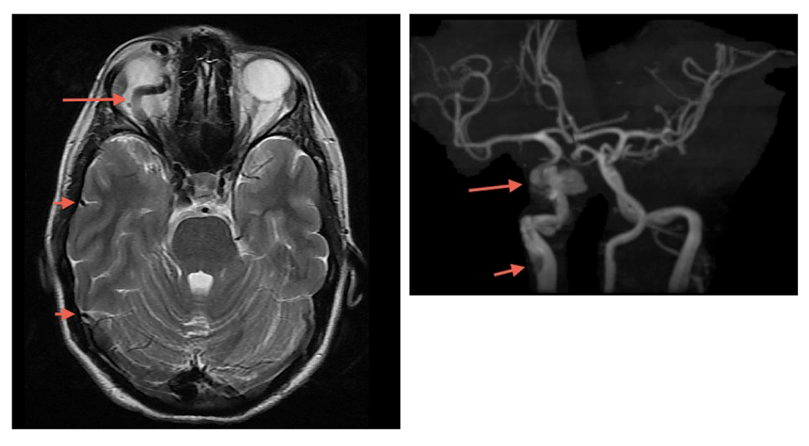
Figure 1. MRI/MRA/MRV of the brain revealed a dilated ophthalmic vein (long arrow), engorged cerebral veins (short arrows), spontaneous dissection of the internal carotid artery at the level of the petrous portion of the temporal bone (short arrow) and a direct carotid cavernous fistula (long arrow).
Figure 2.
Figure 2. The fistula prior to embolization (red arrow). Fistula status post coil embolization (blue arrow).