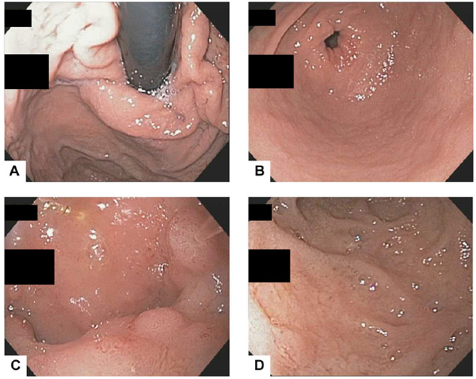
Figure 1. The EGD revealed normal gastric cardia (A) and antrum (B) but mild nodularity in the duodenal bulb (C) and nonspecific duodenitis in the second portion of duodenum (D).
| Journal of Medical Cases, ISSN 1923-4155 print, 1923-4163 online, Open Access |
| Article copyright, the authors; Journal compilation copyright, J Med Cases and Elmer Press Inc |
| Journal website http://www.journalmc.org |
Case Report
Volume 4, Number 3, March 2013, pages 166-169
Isolated Russell Body Duodenitis with Concurrent Helicobacter Pylori Gastritis
Figures
Table
| Author | Age at Diagnosis | Sex | Clinical Features | Endoscopic Features | Pathologic Features | ||
|---|---|---|---|---|---|---|---|
| Stomach | Duodenum | Immunophenotype of Russel body containing plasma cells | |||||
| Savage et al, 2011 | 55 yrs | M | 2-week history abdominal pain; HIV positive with undetectable viral load and CD4 of 514 /mm3; history of lymphoma (5- year remission) | Non-specific gastritis and duodenitis without identifiable mass | Mild chronic gastritis, no Russell bodies, negative for H. pylori | Duodenal mucosa with dense infiltrate of plasma cells in lamina propria that contained numerous Russell bodies, mild peptic duodenitis | CD138+, cytokeratin-, polytypic kappa and lambda |
| Paniz Mondolfi et al, 2012 | 69 yrs | F | Multiple complaints of dysphagia with prior endoscopy showing Schatzki ring, and repeat showing esophagitis and gastroduodenitis; history of Crohn’s disease, cirrhosis, rheumatoid arthritis, morbid obesity with sleeve gastrectomy | Nodule within duodenal bulb | Negative for H. pylori | Enteric-type mucosa with gastric metaplasia and numerous lamina propria plasma cells containing Russell bodies | CD138+, CD68-, polytypic kappa and lambda |
| Current study | 59 yrs | F | Epigastric pain, bloating, and diarrhea of several years duration; history of type II diabetes mellitus, gastroesophageal reflux disease and irritable bowel syndrome, and chronic obstruction pulmonary disease | Nodular mucosa in the duodenum, normal appearing esophagus and stomach | Mild chronic gastritis with patchy activity, no Russell bodies, positive for H. pylori | Duodenal mucosa with dense infiltrate of Russell body containing plasma cells in lamina propria, mild peptic duodenitis | CD138-, CD68-, AE1/AE3-, polytypic kappa and lambda |