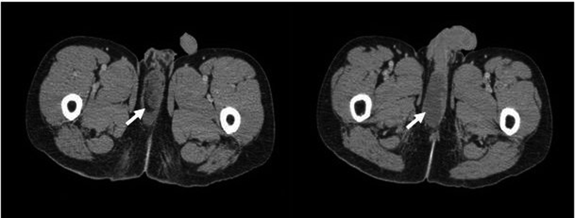
Figure 1. Contrast- enhanced CT of a 36 years old male patient with the complaint of pain and swelling at the right perineal region showed hypodense area (in fluid density) in the perineal region which was at the location of the right corpus spongiosum, between the both corpus cavernosa and at the right portion of the penil root (arrow). (Toshiba Aquilion 64 Detector CT, Istanbul/Turkey. CT Protocol: 50 mAs, 120 kV, 7 mm Slice thickness, 350 mL iodine contrast was used).
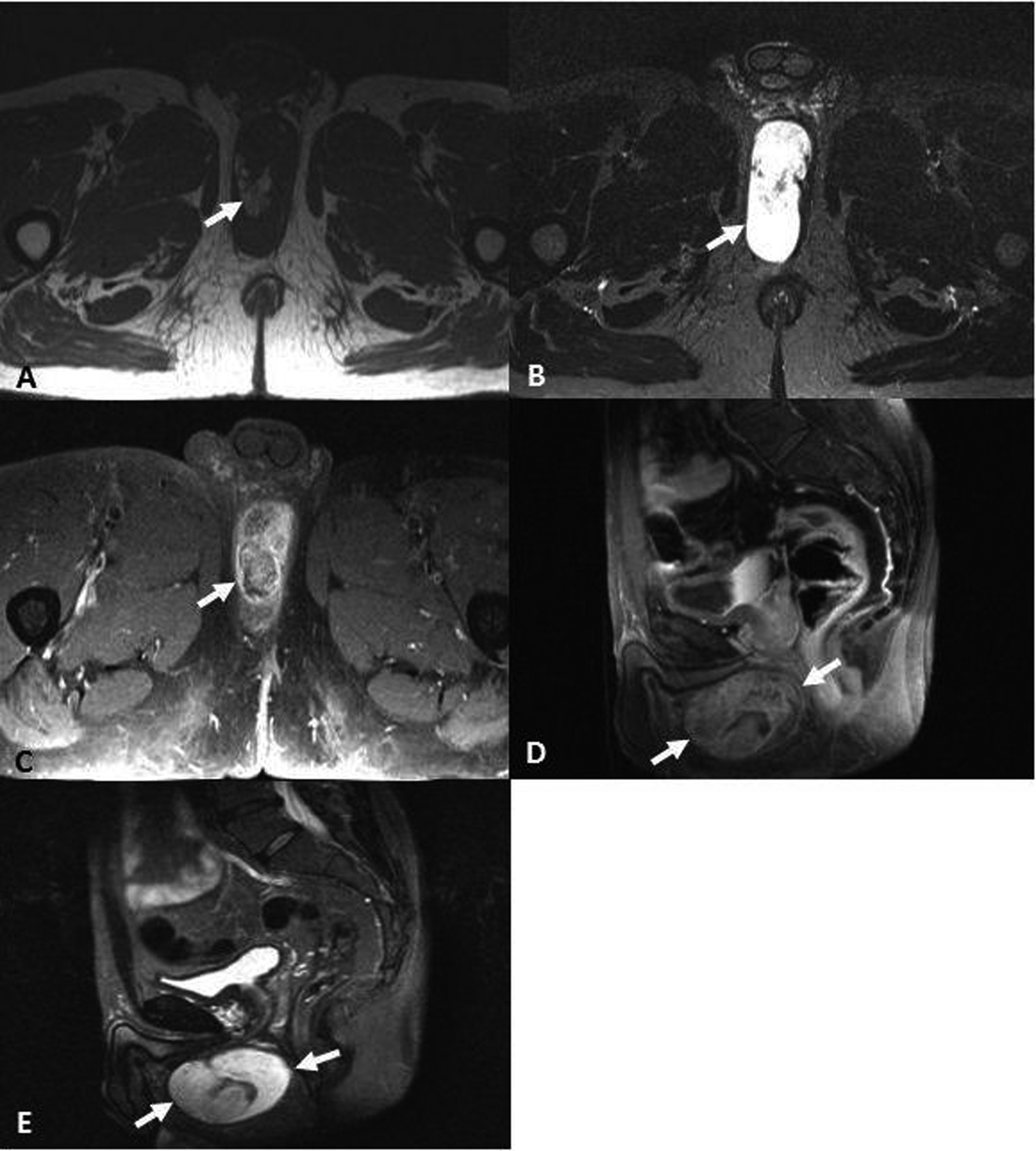
Figure 2. Pelvic MR Imaging of of a 36 years old male patient with the complaint of pain and swelling at the right perineal region revealed a 8 × 6 × 3 cm sized lesion cystic in nature, hyperintense on axial T1-Weighted images (TE: 600, TR: 10) (A), remarkable hyperintense in signal intensity on axial and sagittal STIR sequence TE: 85, TR: 3000) (B, C), heterogenous and mural contrast enhacement pattern on axial and sagittal post-contrast imaging (TE: 635 TR: 15 ), (D, E) was observed. (Philips 1.5 Tesla MR, Istanbul/Turkey. Godalinium contrast material 20 mg was admitted).