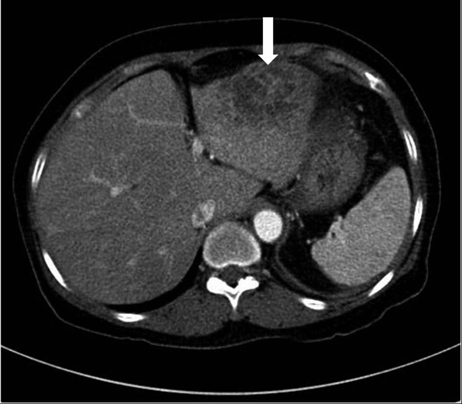
Figure 1. The initial CT scan shows a 5.5 x 3.5 cm, oval shaped, heterogeneously low attenuated lesion in the left lateral segment of the liver (white arrow).
| Journal of Medical Cases, ISSN 1923-4155 print, 1923-4163 online, Open Access |
| Article copyright, the authors; Journal compilation copyright, J Med Cases and Elmer Press Inc |
| Journal website http://www.journalmc.org |
Case Report
Volume 3, Number 6, December 2012, pages 370-372
Pyogenic Liver Abscess With Complicating Intestinal Tuberculosis
Figures
