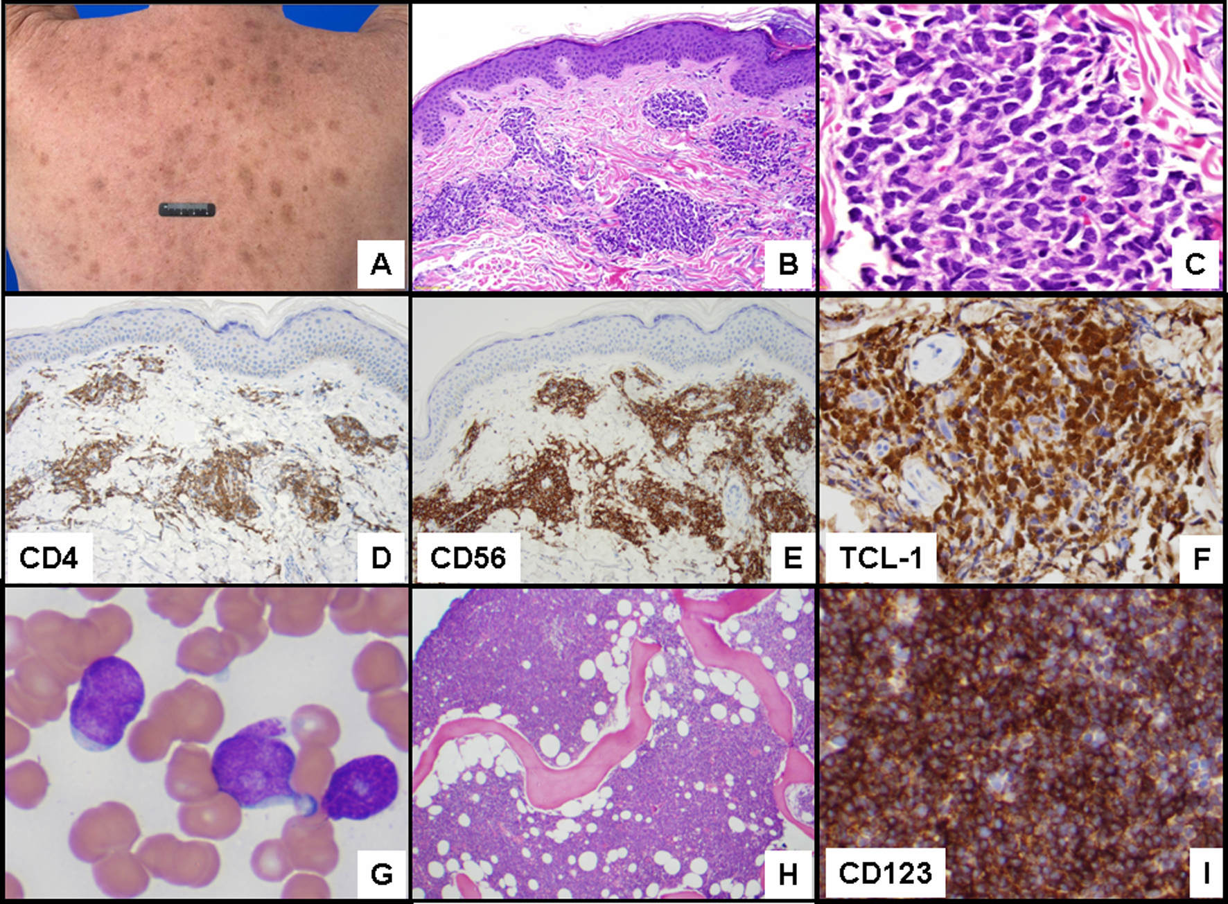
Figure 1. (A) Multiple purple to brown macules and papules on the back. (B) Skin biopsy with neoplastic cells in the dermis distributed in a perivascular and periadnexal pattern (hematoxylin-eosin (H & E) stain, original magnification 100 x). (C) Monotonous infiltrate of blastic appearing neoplastic cells in the dermis (H & E stain, original magnification, 400 x) positive for (D) CD4 (original magnification 100 x), (E) CD56 (original magnification 100 x, and (F) TCL-1 (original magnification 200 x). (G) Peripheral blood smear with circulating blasts (Wright Giemsa, original magnification 1,000 x), (H) and bone marrow with acute leukemia (H & E, original magnification 40 x). (I) Leukemic cells positive for CD123 (original magnification 200 x).