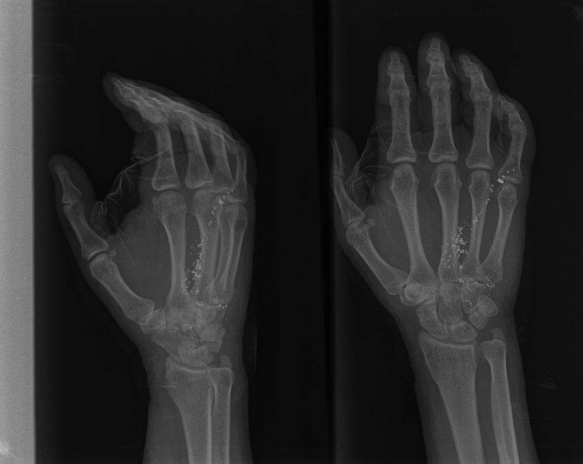
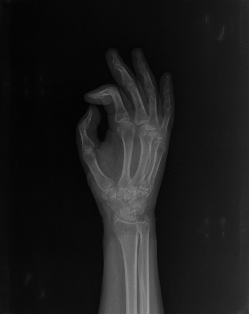
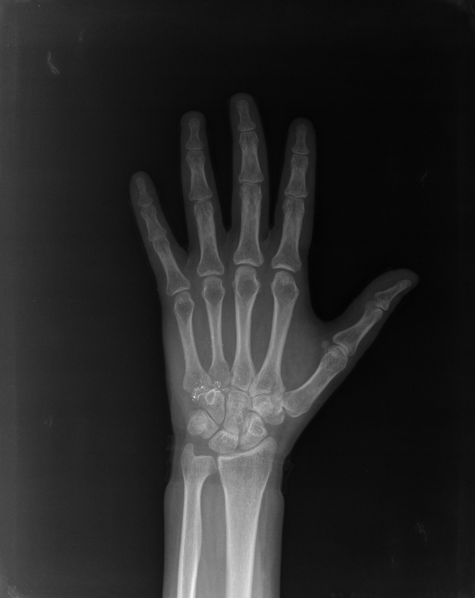
Figure 1. Preoperative lateral and anteroposterior radiographs of the left hand showing multipl scattered metal-density opacities localized in the soft tissues around the metacarpus.
| Journal of Medical Cases, ISSN 1923-4155 print, 1923-4163 online, Open Access |
| Article copyright, the authors; Journal compilation copyright, J Med Cases and Elmer Press Inc |
| Journal website http://www.journalmc.org |
Case Report
Volume 3, Number 6, December 2012, pages 365-369
Administration of Metallic Mercury by an Accidental Puncture in the Hand: A Case Report
Figures
Table
| Mercury Level (blood) µg/L | Mercury Level (24-h urine) µg/d | |
|---|---|---|
| 17.06.2011 | 12.20 | 13.20 |
| 31.06.2011 | 14.30 | 23.30 |
| 16.07.2011 | 24.00 | 33.20 |
| 01.08.2011 | 9.80 | 25.00 |
| 17.08.2011 | 10.80 | 35.00 |
| 15.09.2011 | 11.77 | 85.60 |
| 17.10.2011 | 19.60 | 125.00 |
| 31.10.2011 | 16.80 | 172.00 |
| 21.01.2012 | 9.70 | 58.00 |
| 16.03.2012 | 3.70 | 42.00 |