Journal of Medical Cases, ISSN 1923-4155 print, 1923-4163 online, Open Access
Article copyright, the authors; Journal compilation copyright, J Med Cases and Elmer Press Inc
Journal website http://www.journalmc.org

Case Report

Volume 3, Number 6, December 2012, pages 347-351


Takotsubo Cardiomyopathy and Acute Ischemic Stroke

Figures

Figure 1.
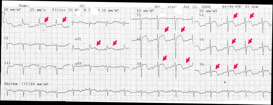
Figure 1. Electrocardiogram performed on admission showed supra ST deviation and inverted T waves in DI, AVL and V2-V6 leads (colored arrows).
Figure 2.
Figure 2. Ventriculography on diastole (a) and systole (b), showing apical ballooning and acinesia and left ventricle basal hypercontractility with compromised systolic function (c). Coronariography (d) showing no stenotic lesions in left and right coronary arteries. LV: Left ventricle, LC: left coronary artery, RC: right coronary artery.
Figure 3.
Figure 3. Cerebral MRI performed at the 7th day-DWI sequence-Showing multiple acute ischemic lesions: large left temporo-parietal and insular ischemic infarct (a), multiple small infarcts in right medial and posterior cerebral arteries (a, b) and small left cerebellar hemispheric infarct (c).