Journal of Medical Cases, ISSN 1923-4155 print, 1923-4163 online, Open Access
Article copyright, the authors; Journal compilation copyright, J Med Cases and Elmer Press Inc
Journal website http://www.journalmc.org

Case Report

Volume 4, Number 1, January 2013, pages 15-18


Pancreatoduodenectomy With Blood Vessel Reconstruction for a Huge Solid Pseudopapillary Tumor of the Pancreas: Report of a Case

Figures

Figure 1.
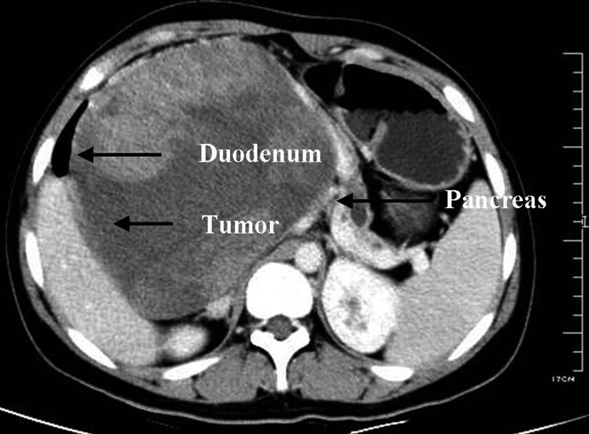
Figure 1. CT scan showing a cystic mass (12.2 cm * 15.4 cm) at the head of the pancreas in a 19-year-old girl with duodenum and pancreas compressed.
Figure 2.
Figure 2. CTA showed that SMA was invaded.
Figure 3.
Figure 3. MRCP showed a space-occupying lesion (15.9 cm * 14.6cm) located in the right upper abdomen, with compressed and displacement of the common bile duct and dilation of pancreatic body and tail duct
Figure 4.
Figure 4. Gross appearance. A. The tumor located in the head of the pancreas with duodenum, pancreas and SMV compressed. B. After reconstruction of blood vessels. C & D. Photograph of gross specimen showing a circumscribed, capsulated, round, gray-purple colored tumor was identified in the pancreatic head which contains varying amounts of necrosis, hemorrhage and cystic change.