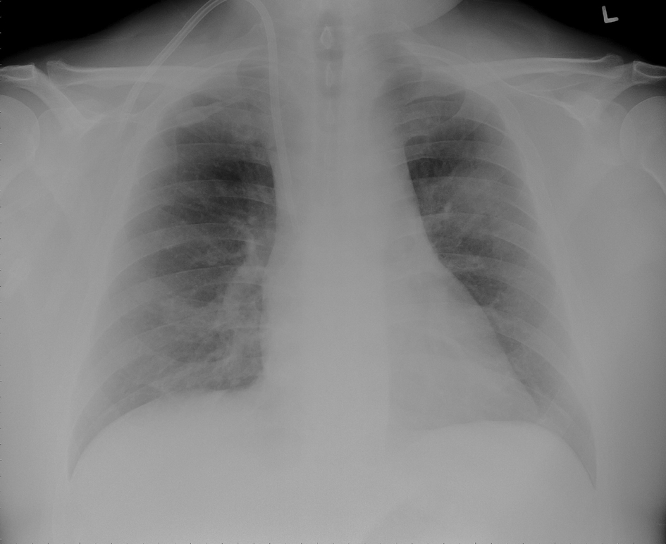
Figure 1. Chest X-ray showing left upper lobe mass like infiltrate and a tunneled ash catheter in the right internal jugular vein.
| Journal of Medical Cases, ISSN 1923-4155 print, 1923-4163 online, Open Access |
| Article copyright, the authors; Journal compilation copyright, J Med Cases and Elmer Press Inc |
| Journal website http://www.journalmc.org |
Case Report
Volume 4, Number 3, March 2013, pages 149-152
Hemodialysis-Catheter Related Septic Pulmonary Emboli in the Absence of Endocarditis or Right Atrial Thrombus
Figures