Journal of Medical Cases, ISSN 1923-4155 print, 1923-4163 online, Open Access
Article copyright, the authors; Journal compilation copyright, J Med Cases and Elmer Press Inc
Journal website http://www.journalmc.org

Case Report

Volume 3, Number 5, October 2012, pages 308-311


Subacute Thyroiditis With Coexisting Papillary Carcinoma Diagnosed by Immediately Repeat Fine Needle Aspiration: A Case Report

Figures

Figure 1.
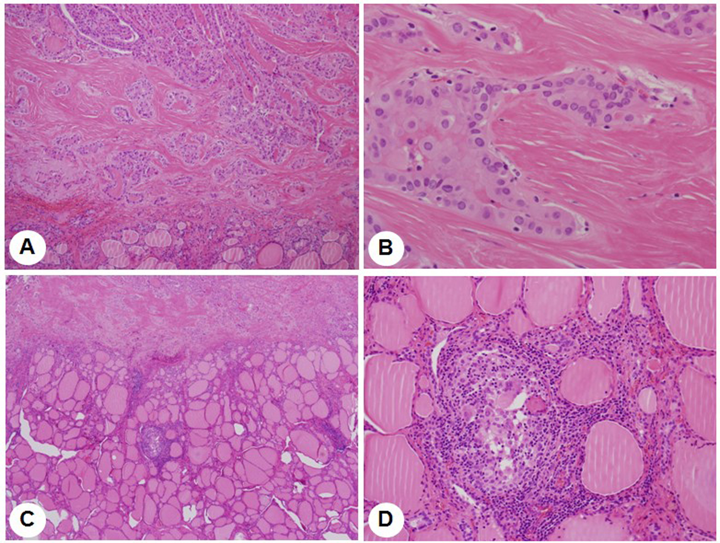
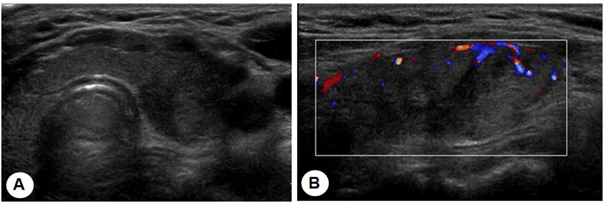
Figure 1. (A) The transverse and longitudinal thyroid sonograms show ill-defined hypoechoic areas in the periphery of both lobes of the thyroid and a 1.2 x 0.8 cm hypoechoic, irregularly-shaped nodule in the left lobe of the thyroid. (B) Color Doppler ultrasonography shows slightly increased vascularity in the hypoechoic areas.
Figure 2.
Figure 2. Tc-99m scintigraphy with non-visualization of the thyroid gland.
Figure 3.
Figure 3. Pathological finding reveals a papillary carcinoma (A, x 40, and B, x 400, H-E stain) and a granuloma surrounded by small lymphocytes in the thyroid parenchyme (C, x 40, and D, x 200, H-E stain).