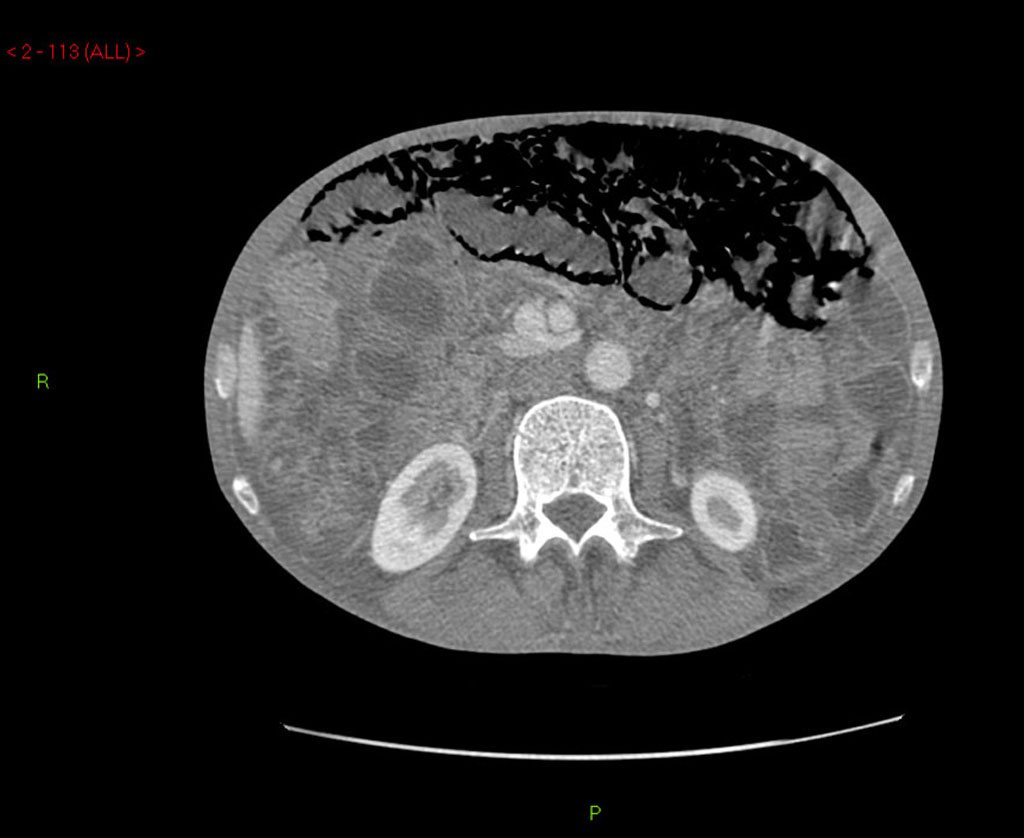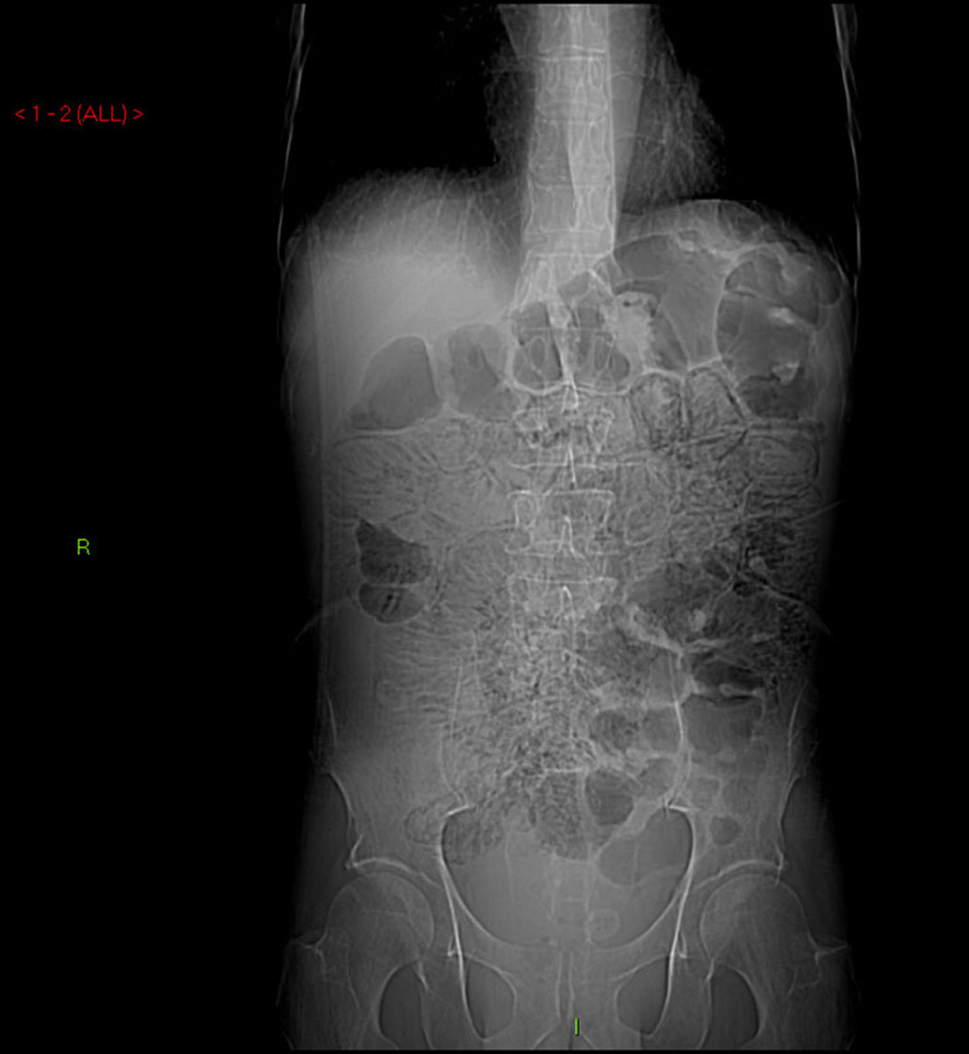
Figure 1. Abdominal X-ray demonstrating extensive pneumatosis intestinalis (PI).
| Journal of Medical Cases, ISSN 1923-4155 print, 1923-4163 online, Open Access |
| Article copyright, the authors; Journal compilation copyright, J Med Cases and Elmer Press Inc |
| Journal website http://www.journalmc.org |
Case Report
Volume 2, Number 2, April 2011, pages 39-43
Extensive Pneumatosis Intestinalis in Association With Celiac Disease: A Case Report
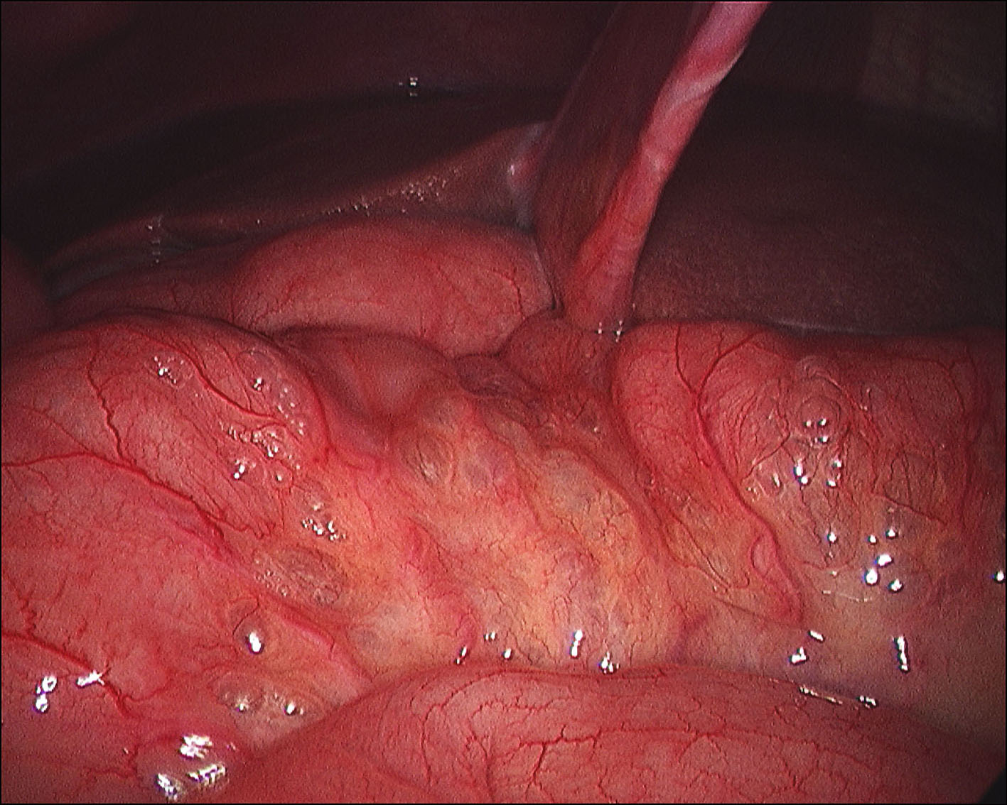
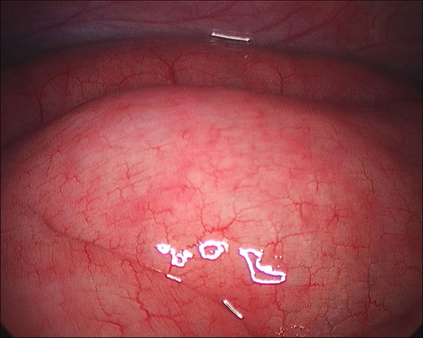
Figures