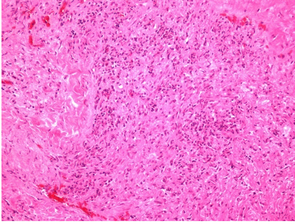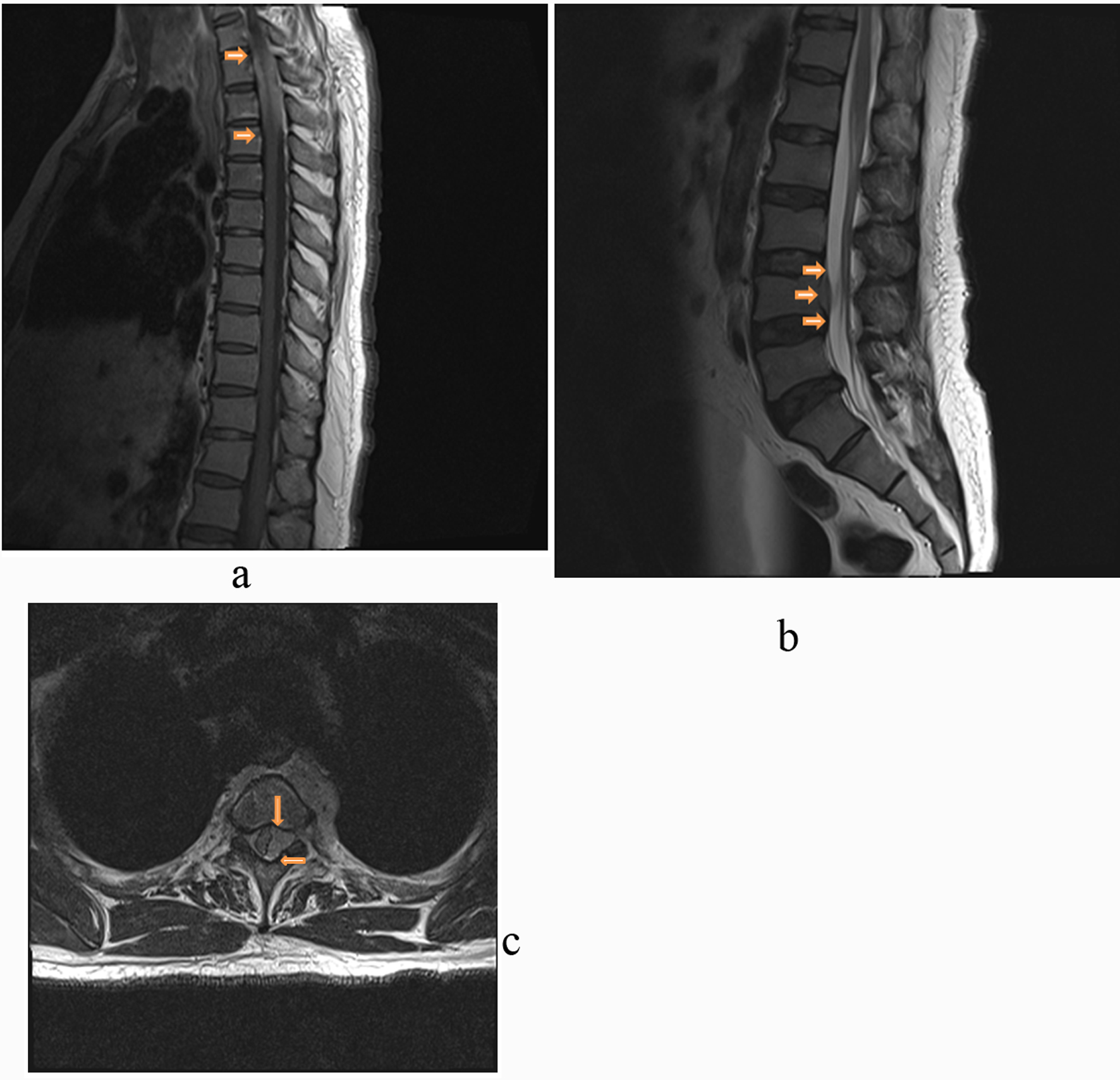
Figure 1. Hypertrophic pachymeningitis shown on MRI of spine with gadolinium contrast. A) Prominent abnormal enhancement infiltrated epidural space from T1 to T3, shown in the T1-weighted post-gadolinium sagittal image of thoracic spine. B) Prominent abnormal enhancement at ventral side at L4, shown in the T2-weighted post-gadolinium sagittal image of lumbar spine. C). Abnormal soft tissue on the left lateral side with mass effect on spinal cord, shown in the T2-weghted post-gadolinium transverse image at the thoracic T3. Arrows point to the thickened dura.