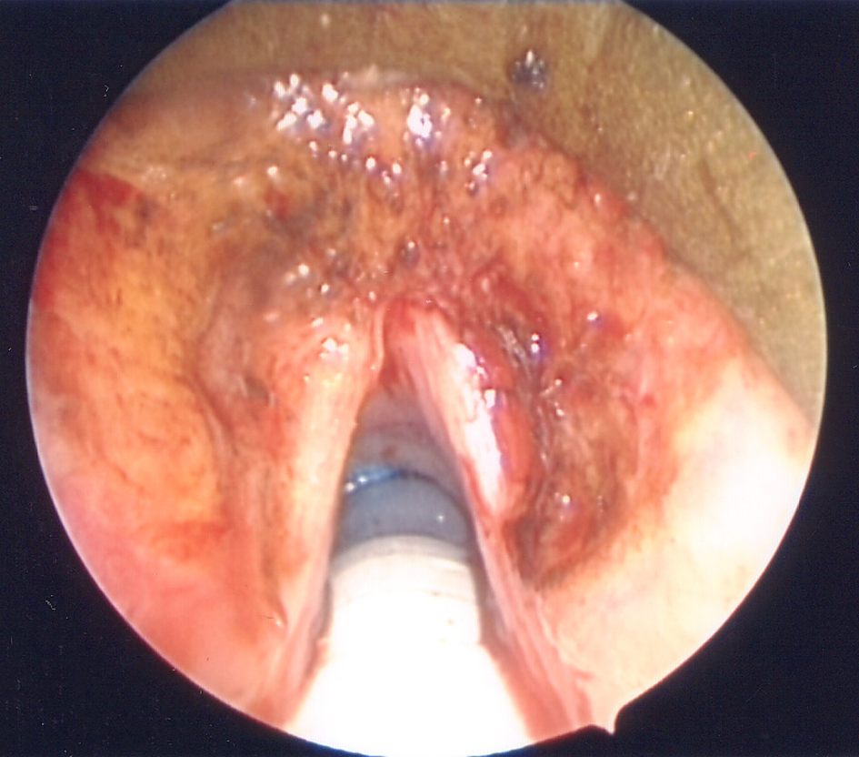
Figure 1. Image of the patient’s original laryngovideostroboscopy. Note the smooth submucosal nodularity of the anterior vestibular folds and ventricles as well as inflammation of the left vocal fold due to amyloid infiltration.
| Journal of Medical Cases, ISSN 1923-4155 print, 1923-4163 online, Open Access |
| Article copyright, the authors; Journal compilation copyright, J Med Cases and Elmer Press Inc |
| Journal website http://www.journalmc.org |
Case Report
Volume 4, Number 1, January 2013, pages 46-48
Recurrent Primary Laryngeal Amyloidosis in a 36 Year-Old-Woman
Figures