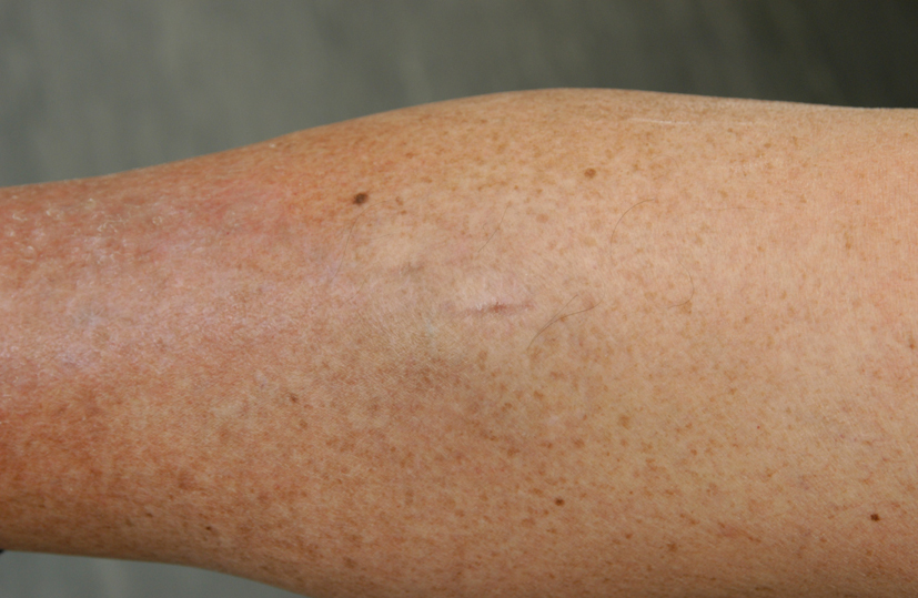
Figure 1. Pre-operative photograph of lesion showing raised lesion on proximal third of right tibia.
| Journal of Medical Cases, ISSN 1923-4155 print, 1923-4163 online, Open Access |
| Article copyright, the authors; Journal compilation copyright, J Med Cases and Elmer Press Inc |
| Journal website http://www.journalmc.org |
Case Report
Volume 3, Number 6, December 2012, pages 334-336
Pleomorphic Hyalinising Angiectatic Tumour: A Tumour Diagnosis of Suspicion
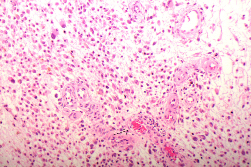
Figures