Journal of Medical Cases, ISSN 1923-4155 print, 1923-4163 online, Open Access
Article copyright, the authors; Journal compilation copyright, J Med Cases and Elmer Press Inc
Journal website http://www.journalmc.org

Case Report

Volume 3, Number 3, June 2012, pages 174-177


Septic Thrombophlebitis Complicating a Peripherally Inserted Central Catheter

Figures

Figure 1.
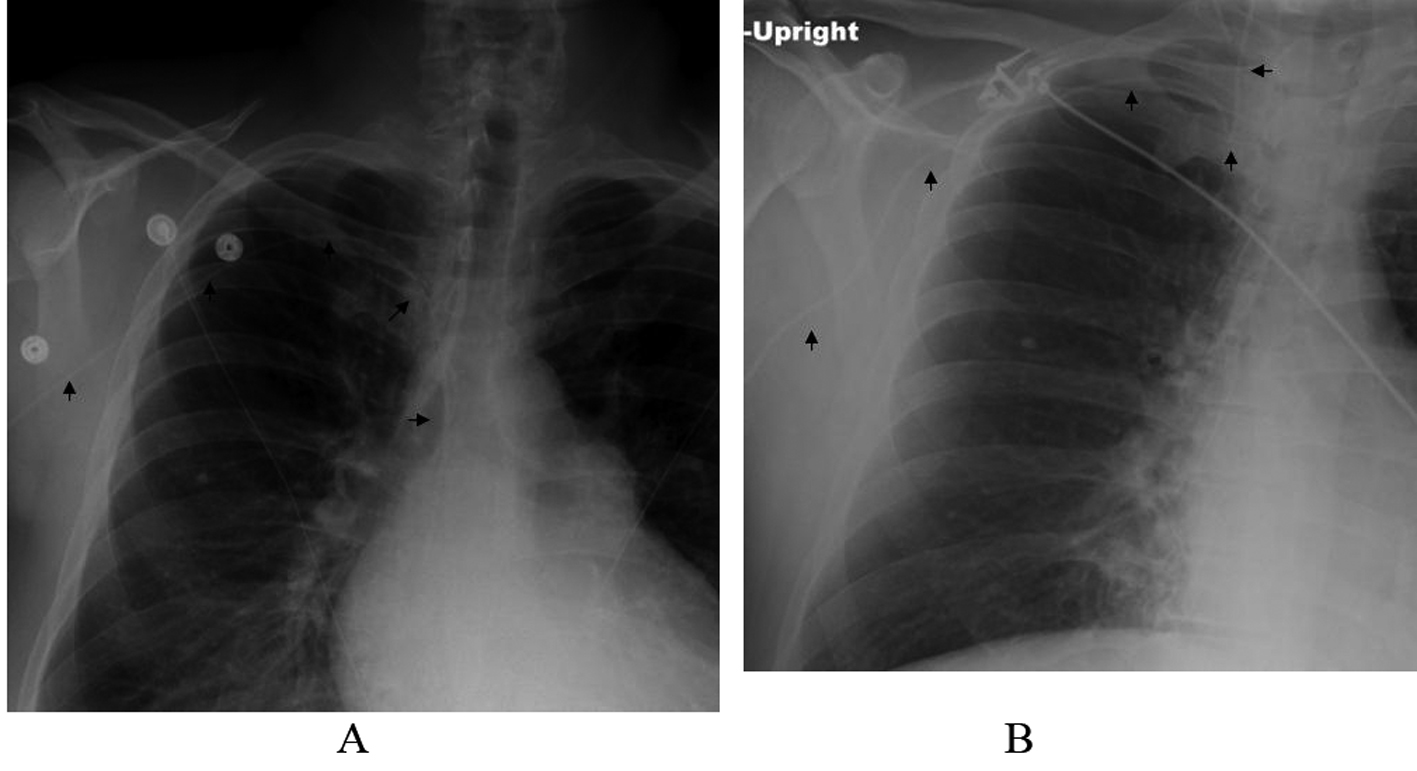
Figure 1. A, Portable chest x-ray after placing the PICC showing a correct positioning (arrows). B, Portable chest x-ray 3 months later showing displacement of the PICC into the right internal jugular vein (arrows).
Figure 2.
Figure 2. A, Non-contrast CT scan of the neck showing admixture of air and fluid in the right internal jugular vein. B, Non-contrast CT scan of the chest showing extension of the thrombophlebitis to the right brachicephalic vein and superior vena cava. C, Non-contrast CT scan of the neck showing air in the internal jugular vein (short white arrow), common facial vein (long white arrow), external facial vein (long black arrow) and internal facial vein (short black arrow).
Figure 3.
Figure 3. A, CT scan of the neck and chest with contrast showing thrombosis of the right internal jugular vein, right subclavian vein, right brachiocephalic and superior vena cava with a small residual pocket of air inferior to the jugular foramen (arrow). B, CT scan of the neck with contrast showing thrombosis of the internal jugular vein (white arrow), external facial vein (short black arrow) and internal facial vein (long black arrow).