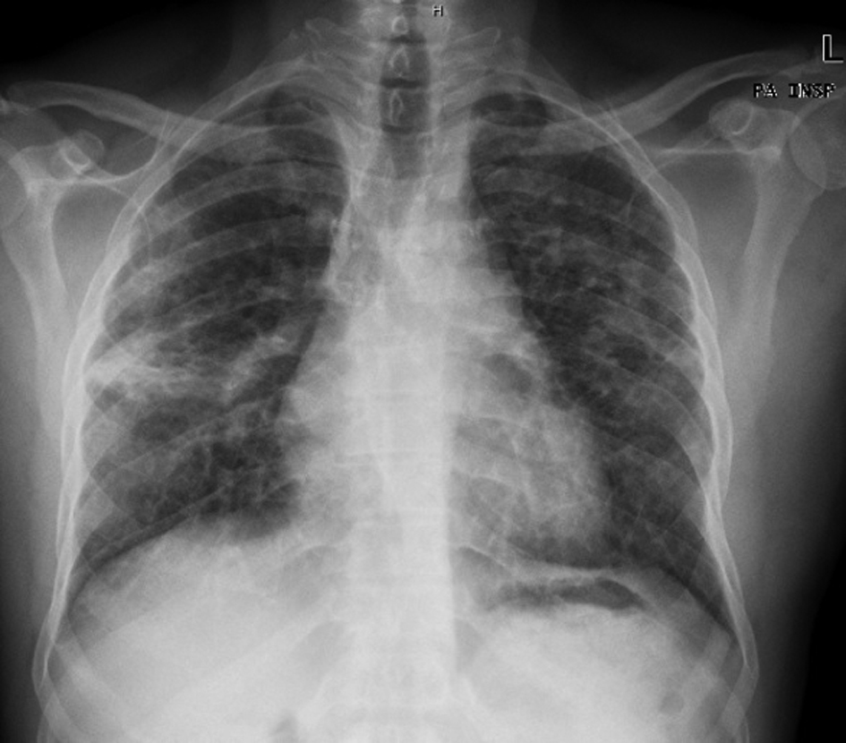
Figure 1. Chest X-ray showing opacity of a fibrotic appearance in both lung fields, most exuberant in the middle level of the right lung field.
| Journal of Medical Cases, ISSN 1923-4155 print, 1923-4163 online, Open Access |
| Article copyright, the authors; Journal compilation copyright, J Med Cases and Elmer Press Inc |
| Journal website http://www.journalmc.org |
Case Report
Volume 3, Number 2, April 2012, pages 155-159
Hypersensitivity Pneumonitis: A Clinical Case
Figures