Journal of Medical Cases, ISSN 1923-4155 print, 1923-4163 online, Open Access
Article copyright, the authors; Journal compilation copyright, J Med Cases and Elmer Press Inc
Journal website http://www.journalmc.org

Case Report

Volume 3, Number 2, April 2012, pages 135-139


Hemophilus Parainfluenzae Pneumonia With Huge IVC Thrombosis and Pulmonary Embolism in a Young Immunocompetent Male

Figures

Figure 1.
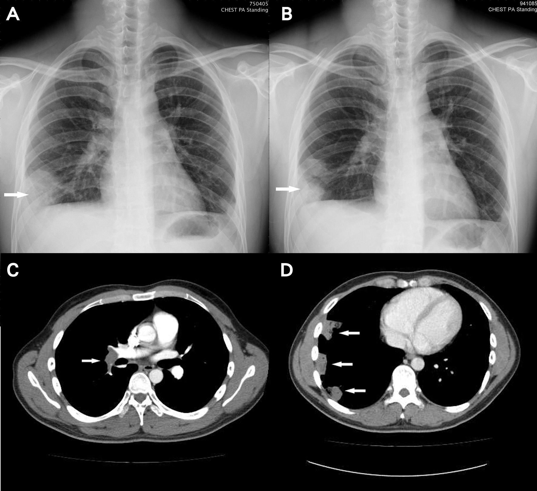
Figure 1. A: Chest radiograph on admission showing air space opacification over right lower lobe (arrow); B: Chest radiograph after empiric antibiotics treatment showing separated consolidative patch over right lower lobe (arrow) with slight resolution; C: CT angiogram showing pulmonary embolism of right main and descending arteries (arrow); D: chest CT demonstrating peripheral patches and consolidation in the right lower lung (arrow) corresponding to the consolidative patch seen on plain film.
Figure 2.
Figure 2. CT angiogram showing abnormal filling defect over right main pulmonary artery and right descending pulmonary artery (arrow) compatible with pulmonary embolism of right main and descending pulmonary arteries.
Figure 3.
Figure 3. CT angiogram showing complete thrombosis of left femoral to common iliac veins, and partial thrombosis of right common iliac vein to infrarenal IVC (lower arrow).
Figure 4.
Figure 4. A: Venocavogram showing a filling defect in IVC (arrow) and right common iliac vein, and non-opacification of left iliac vein; B: A COOK Celect IVC filter deployed at infrarenal IVC (arrow).