Journal of Medical Cases, ISSN 1923-4155 print, 1923-4163 online, Open Access
Article copyright, the authors; Journal compilation copyright, J Med Cases and Elmer Press Inc
Journal website http://www.journalmc.org

Case Report

Volume 3, Number 2, April 2012, pages 106-109


An Unusual Case of Bilateral Submandibular Sialolithiasis

Figures

Figure 1.
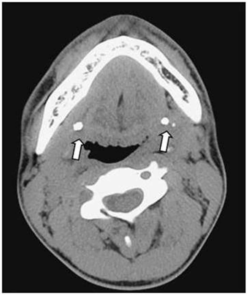
Figure 1. Axial computed tomography scans demonstrating bilateral calcified densities in the submandibular gland (arrows).
Figure 2.
Figure 2. Surgical approach to the stones in the warton’s duct or the hilum of the gland. A portion of sublingual gland overlying the proximal duct was removed to expose the proximal duct(black arrow) and lingual nerve(white arrow). The lingual nerve crossing the wharton’s duct was retracted to the inside (A, B). The stone in the hilum of submandibular gland was exposed (arrow head) by pushing upward the subman¬dibular triangle using the fingers and incision made on the upper area of the Wharton’s duct where the stone was palpated (C, D).
Figure 3.
Figure 3. Two stones smaller than 1cm in the hilum of both submandibular gland, and tiny one stone in the parenchyma of submandibular gland were removed.