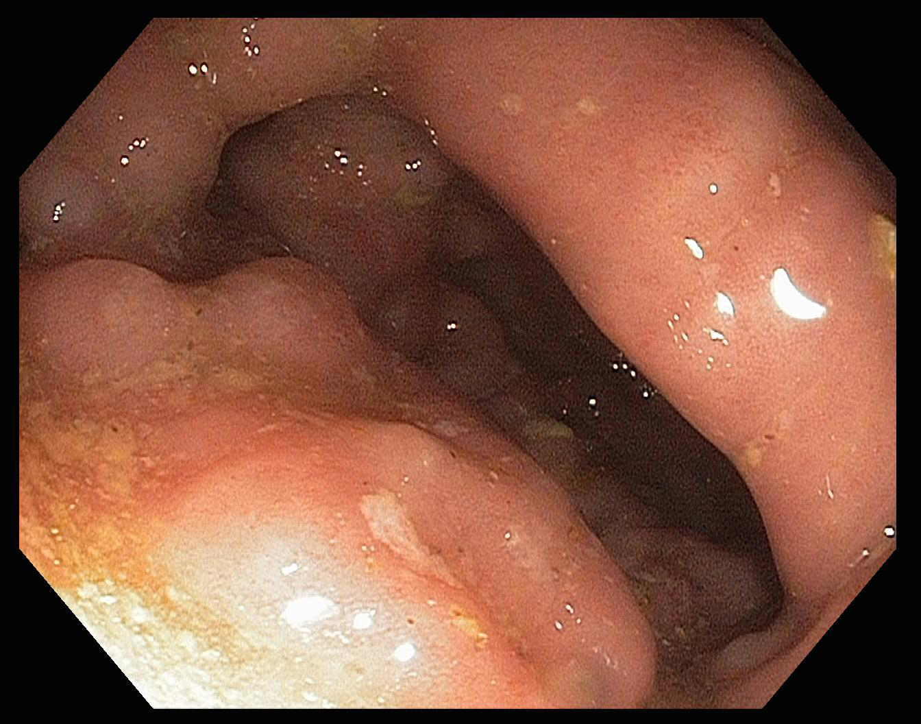
Figure 1. Colonoscopy Image. There are markedly edematous folds in most of the right colon with multiple small superficial erosions.
| Journal of Medical Cases, ISSN 1923-4155 print, 1923-4163 online, Open Access |
| Article copyright, the authors; Journal compilation copyright, J Med Cases and Elmer Press Inc |
| Journal website http://www.journalmc.org |
Case Report
Volume 3, Number 1, February 2012, pages 49-53
Isolated Colonic Metastasis From Primary Invasive Ductal Breast Carcinoma: Role of Tumor Marker in Early Diagnosis
Figures
Table
| References | Age | Patients | Time since primary (year) | Histology | Morphology | Tumor markers | Signs and symptoms |
|---|---|---|---|---|---|---|---|
| Taal et al [3] | 56 | 17 | 4.4 | Ductal/lobular | Stricture | NA | Varied |
| Theraux et al [4] | 69 | 1 | 28 | Ductal | Stricture | NA | Obstruction |
| Feng et al [5] | 49 | 1 | 2 | Ductal | Colonic erosion | NA | Abdominal Pain |
| Alves De lima et al [6] | 74 | 1 | 7 | Lobular | Stricture | Normal | Anemia |
| Koustsomanias et al [7] | 65 | 1 | 4 | Ductal | NA | NA | Anemia |
| Uygun K et al [8] | 43 | 1 | 4.9 | Ductal/lobular | Stricture | Normal | Change in bowel habits |
| Rabau et al [9] | 53 | 1 | 7 | Lobular | Stricture | NA | Obstruction |
| Schwarz et al [10] | 75 | 1 | 0.25 | Ductal/lobular | Mucosal Ulcer | NA | Abdominal mass |
| Okido et al [11] | 48 | 1 | 5 | Ductal/lobular | Stricture | CEA 52.2 CA 15-3 normal | Abdominal mass |
| Vaidya et al [12] | 56 | 1 | 5 | Ductal | Polypoid tumor | NA | Change in bowel habits |
| Yokota et al [13] | 57 | 1 | 10 | Ductal | Stricture | NA | Asymptomatic |