Journal of Medical Cases, ISSN 1923-4155 print, 1923-4163 online, Open Access
Article copyright, the authors; Journal compilation copyright, J Med Cases and Elmer Press Inc
Journal website https://www.journalmc.org

Case Report

Volume 14, Number 12, December 2023, pages 413-418


Catheter Ablation of Left Ventricular Summit Ectopies in Left Ventricular Noncompaction

Figures

Figure 1.
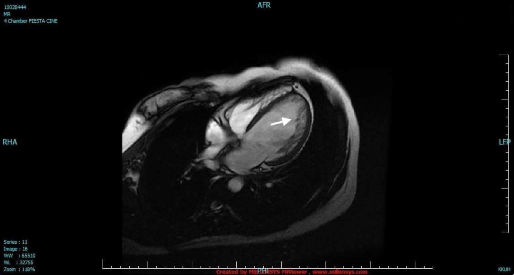
Figure 1. Cardiac magnetic resonance imaging. The LV was dilated with two distinct myocardial layers: a compact layer and a noncompact layer with prominent trabeculations and deep intertrabecular recesses in the lateral wall from base to apex (arrow). The LV systolic function was moderately to severely depressed. LV: left ventricle.
Figure 2.
Figure 2. Baseline electrocardiogram. The rhythm was sinus with complete left bundle branch block. There were monomorphic premature ventricular complexes (right bundle branch block morphology, inferior axis, QS in lead I and aVL, QRS duration of 138 ms) in bigeminal pattern.
Figure 3.
Figure 3. Intracardiac electrogram. The earliest local activation time was recorded in the LV summit. The electrogram in the successful site was preceded by a small presystolic potential by 20 ms (arrow). Aortic valve closure (VC) artifacts were also recorded. LV: left ventricle.
Figure 4.
Figure 4. Ablation site (left anterior oblique and cranial views). Ablation was delivered in the site with the earliest local activation time and presence of presystolic potential. RCC: right coronary cusp; LCC: left coronary cusp; LVOT: left ventricular outflow tract; LV: left ventricle.