Figures
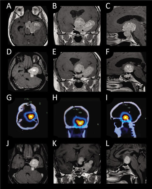
Figure 1. Preoperative T1- weighed MRI with contrast enhancement (A: axial; B: coronal; C: sagittal.). Post operative T1-weighed MRI with contrast enhancement showing resection of the intrasellar portion of the tumor (D: axial; E: coronal; F: sagittal.). Octreoscan images shows lesion (G: axial; H: coronal; I: sagittal.). T1-weighted MRI with contrast enhancement scans 3 months after surgery and Octreotide treatment shows 20% shrinkage of tumor size. (J: axial; K: coronal; L: sagittal)
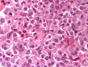
Figure 2. No major cellular and nuclear pleomorphism was seen and mitotic figures were not obvious (Original magnification × 400).
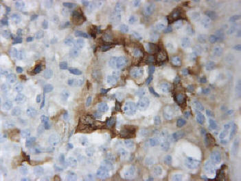
Figure 3. Immuno-positivity for GH in several unevenly distributed adenoma cells (original magnification × 400).
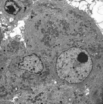
Figure 4. Typical tumor cells having large nucleus, prominent dense nucleolus, but very little heterochromatin (× 7000).
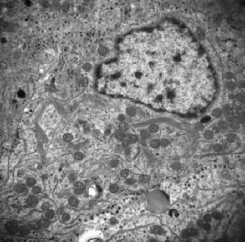
Figure 5. Electron micrograph depicting tumor cells containing collapsed inactive Golgi membrane, but no fibrous body was noted in Golgi area (×15000).