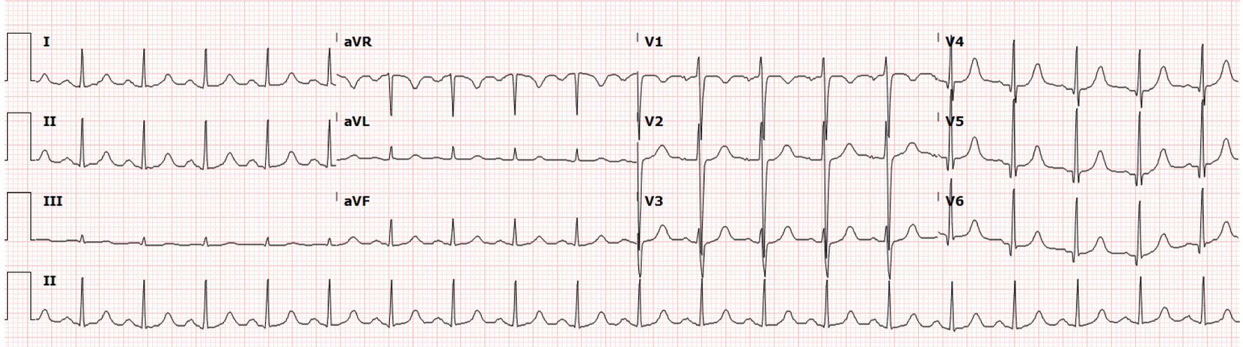
Figure 1. Electrocardiogram at time of presentation to the emergency department.
| Journal of Medical Cases, ISSN 1923-4155 print, 1923-4163 online, Open Access |
| Article copyright, the authors; Journal compilation copyright, J Med Cases and Elmer Press Inc |
| Journal website https://www.journalmc.org |
Case Report
Volume 14, Number 11, November 2023, pages 362-368
Percutaneous Intracardiac Mass Extraction in High Surgical-Risk Patients
Figures
Table
| Device | Aspiration uses | Contraindications | Advantages | Disadvantages |
|---|---|---|---|---|
| ECMO: extracorporeal membrane oxygenation; IVC: inferior vena cava; v-v: veno-venous. | ||||
| AngioVac system | Intracardiac masses; Pulmonary emboli | Severe vascular disease; Chronic thrombi or adherent masses | Minimal blood loss (blood is returned through v-v ECMO) | ECMO causes increased procedural time and cost |
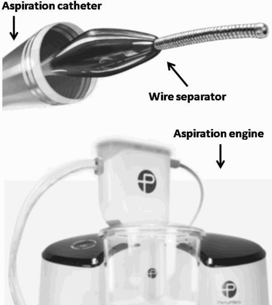
| Penumbra Lightning Aspiration System | Intracardiac masses; Pulmonary emboli; Peripheral thrombi | Pre-existing vascular injuries | ECMO not required | Blood loss is expected during the procedure |
| Inari Medical Device System | Pulmonary emboli; Venous thrombi | IVC filter or venous stents; Small vessels | ECMO not required | Thrombus can be dislodged |