Journal of Medical Cases, ISSN 1923-4155 print, 1923-4163 online, Open Access
Article copyright, the authors; Journal compilation copyright, J Med Cases and Elmer Press Inc
Journal website https://www.journalmc.org

Case Report

Volume 14, Number 12, December 2023, pages 405-412


Idiopathic Hypertrophic Spinal Pachymeningitis

Figures

Figure 1.
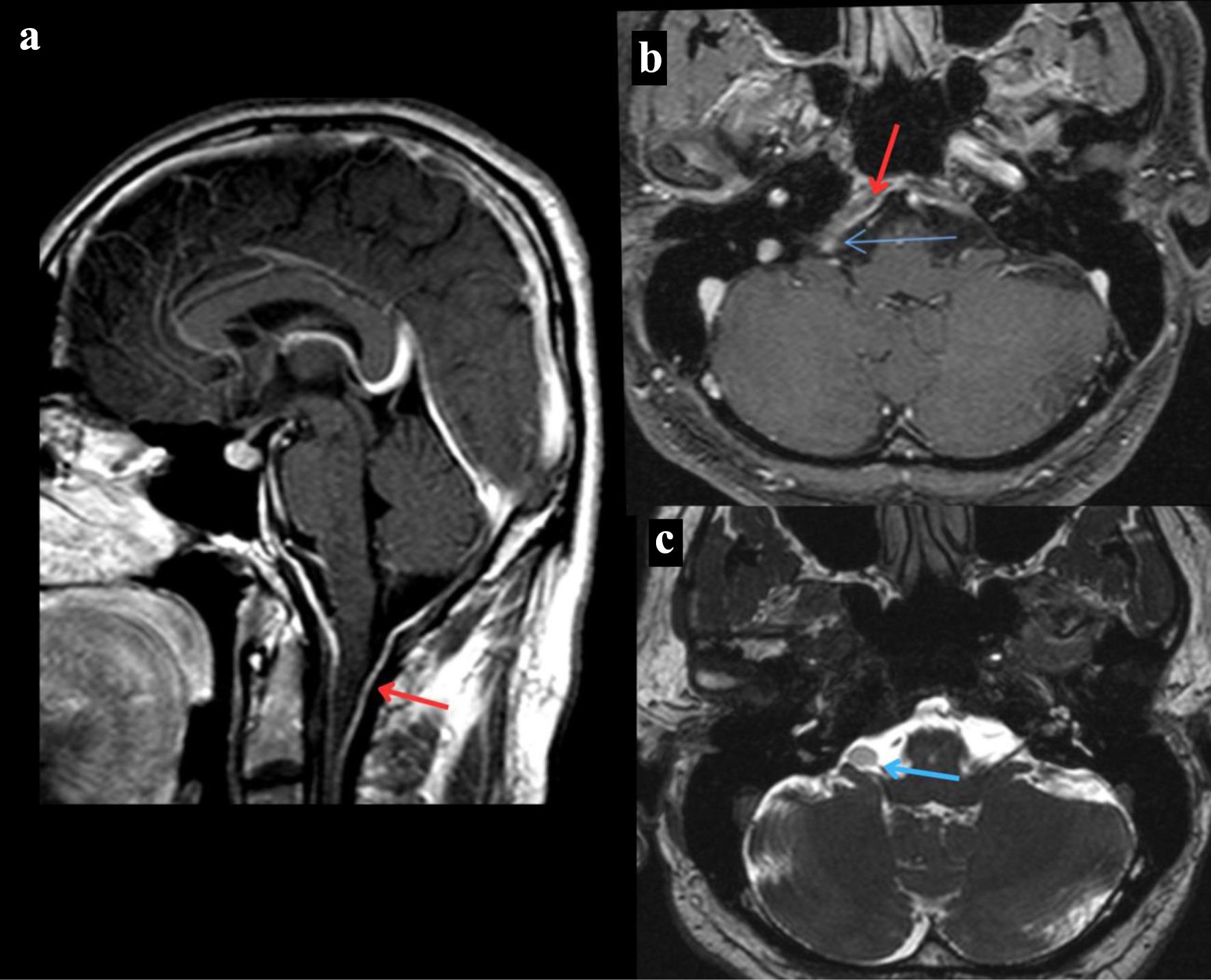
Figure 1. MRI of the brain. (a) T1-weighted MRI of the brain sagittal section post-contrast that reveals dural thickening of the posterior fossa indicated by the red arrow. (b) T1-weighted MRI of the brain axial section post-contrast that reveals a right cerebellopontine angle mass revealed by the blue arrow and dural thickening of the posterior fossa indicated by the red arrow. (c) T2-weighted MRI of the brain axial section that reveals a right cerebellopontine angle mass revealed by the blue arrow. MRI: magnetic resonance imaging.
Figure 2.
Figure 2. MRI cervical spine. (a, b) T1-weighted MRI spine sagittal section without and with contrast showing cervical spine dural thickening with narrowing of cord respectively. (c, d) T1-weighted MRI spine without and with contrast axial section showing narrowing of cord respectively. MRI: magnetic resonance imaging.
Figure 3.
Figure 3. MRI cervical spine. Follow-up T1-weighted MRI with contrast showing decreased meningeal enhancement and narrowing. (a) Sagittal view. (b) Axial view. MRI: magnetic resonance imaging.

Tables

Table 1. CSF Analysis
 
2020201520142013Reference range
aAbnormal results. Significant increase in CSF protein at the time the patient presented with neurologic symptoms is shown. AFB: acid-fast bacilli; CSF: cerebrospinal fluid.
Protein (mg/dL)1,620a263a243a257a15 - 45
Glucose (mmol/L)4952545440 - 85
White cell count (cells/µL)188a, lymphocyte predominance46a, lymphocyte predominanceNot availableNot available0 - 5
Red blood cell count (cells/µL)103aNot availableNot availableNot available0 - 10
AFB cultureNegativeNot availableNot availableNot availableNegative


Table 2. Causes of Hypertrophic Pachymeningitis
 
ANCA: antineutrophil cytoplasmic antibody.
Inflammatory/autoimmune [23-25]Idiopathic (67%)
ANCA-associated vasculitis (15%)
IgG4-related disease
Sarcoidosis
Rheumatoid arthritis
Sjogren syndrome
Systemic lupus erythematosus
Giant-cell arteritis
Infectious [24]Tuberculous meningitis (8%)
Viral meningitis (6%)
Bacterial meningitis (4%)
Neoplastic [24]Lymphoma
Meningioma
Histiocytosis
Carcinomatous meningitis
Other [24]Previous surgical procedure
Cerebrospinal fluid hypotension


Table 3. Outcomes of IHSP With Different Clinical Variables
 
Clinical variableImprovedPartially improvedNot improvedTotal
aInitial intervention was considered in type of management. CRP: C-reactive protein; CSF: cerebrospinal fluid; ESR: erythrocytic sedimentation rate; IHSP: idiopathic hypertrophic spinal pachymeningitis; RA: rheumatoid arthritis; UMN: upper motor neuron.
Course of disease
  Acute (≤ 24 h)1 (25%)1 (25%)2 (50%)4
  Progressive (> 24 h)10 (59%)5 (29%)2 (12%)17
Clinical features
  UMN signs5 (50%)1 (10%)4 (40%)10
CSF findings
  Elevated proteins and leukocyte4 (80%)01 (20%)5
Inflammatory markers
  Elevated ESR, CRP and RA3 (37.5%)3 (37.5%)2 (25%)8
No. of spinal segments
  ≥ 65 (45.4%)4 (36.3%)2 (18.1%)11
  ≤ 56 (60%)2 (20%)2 (20%)10
Type of managementa
  Surgical3 (75%)1 (25%)04
  Conservative4 (100%)004
  Both surgical and conservative4 (30.7%)5 (38.5%)4 (30.7%)13
Location of IHSP
  Anterior and posterior1 (33.3%)2 (66.6%)03
  Anterior3 (75%)01 (25%)4
  Posterior2 (33.3%)2 (33.3%)2 (33.3%)6
  Circumferential4 (57.1%)2 (28.5%)1 (14.2%)7