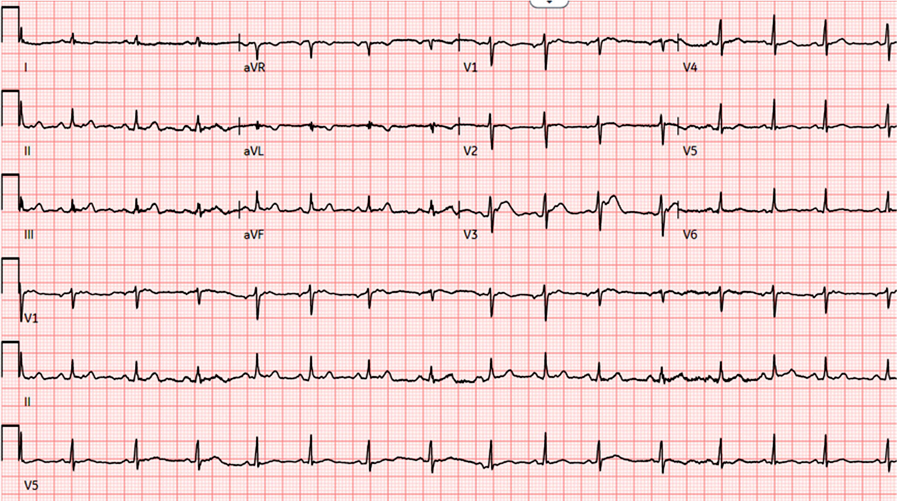
Figure 1. EKG shows low voltage QRS complexes and electrical alternans. EKG: electrocardiogram.
| Journal of Medical Cases, ISSN 1923-4155 print, 1923-4163 online, Open Access |
| Article copyright, the authors; Journal compilation copyright, J Med Cases and Elmer Press Inc |
| Journal website https://www.journalmc.org |
Case Report
Volume 14, Number 8, August 2023, pages 271-276
Tuberculous Pericarditis Presenting as Cardiac Tamponade: Role of Echocardiography
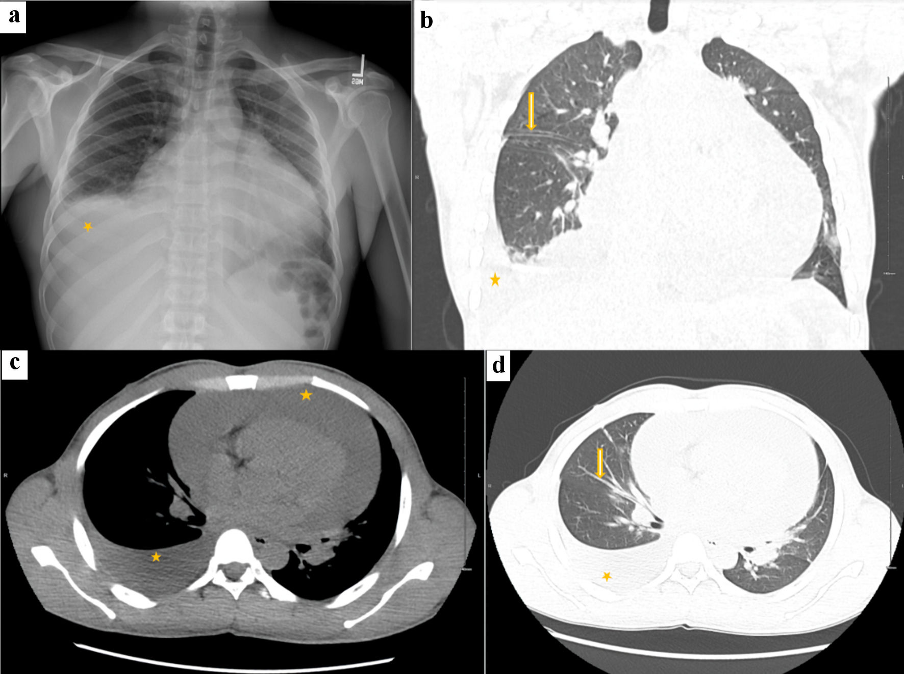
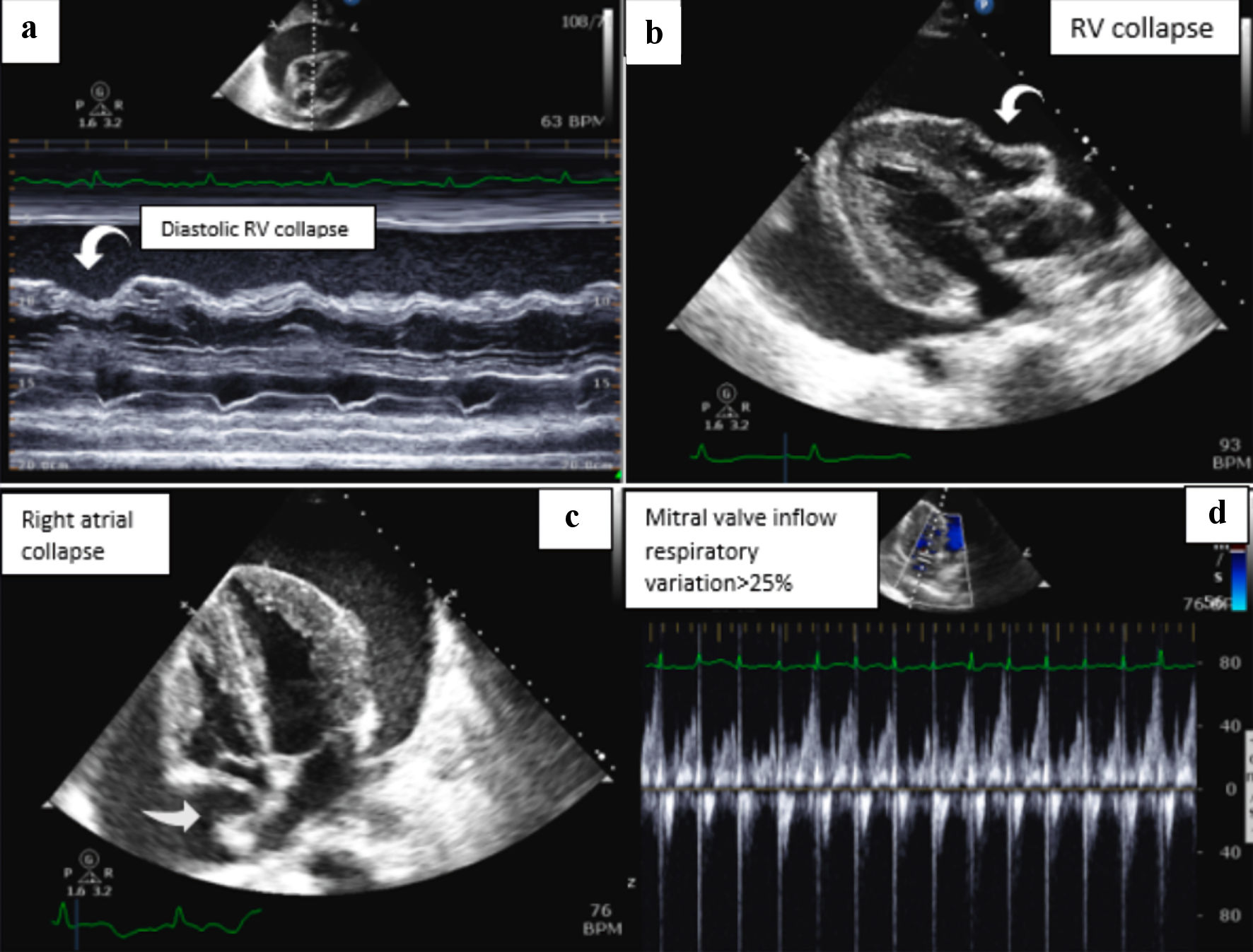
Figures
Table
| Laboratory test | Result | Reference range |
|---|---|---|
| BNP: B-type natriuretic peptide. | ||
| White blood cell counts | 5.70 | 4.50 - 10.90 |
| Hemoglobin | 13.1 g/dL | 12.0 - 16.0 g/dL |
| Platelet count | 327,000/µL | 150,000 - 400,000/µL |
| Aspartate transaminase | 49 U/L | 10 - 35 U/L |
| Alanine transaminase | 77 U/L | 0 - 31 U/L |
| Blood urea nitrogen (BUN) | 15.0 mg/dL | 8.0 - 23.0 mg/dL |
| Creatinine | 1.03 mg/dL | 0.50 - 0.90 mg/dL |
| Pro-BNP | 314 pg/mL | < 125 pg/mL |
| Erythrocyte sedimentation rate (ESR) | 48 mm/h | 0 - 20 mm/h |
| C-reactive protein | 12.20 mg/L | 0 - 3.0 mg/L |
| Troponin T | 0.010 ng/L | < 0.010 ng/L |