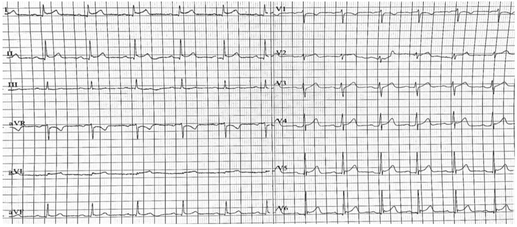
Figure 1. Resting 12-lead electrocardiogram demonstrating sinus rhythm with concave ST-segment elevation in I, II, V4-6.
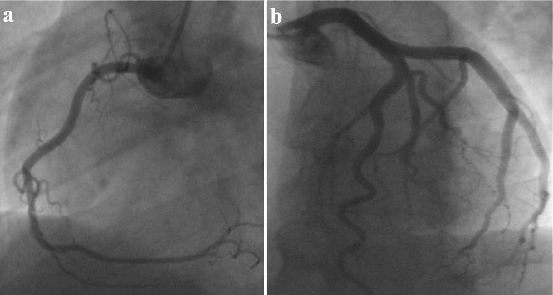
Figure 2. Coronary angiogram revealing normal coronaries, free of significant disease. (a) LAO view of RCA. (b) LAO cranial view of LCA. LAO: left anterior oblique; RCA: right coronary artery; LCA: left coronary artery.
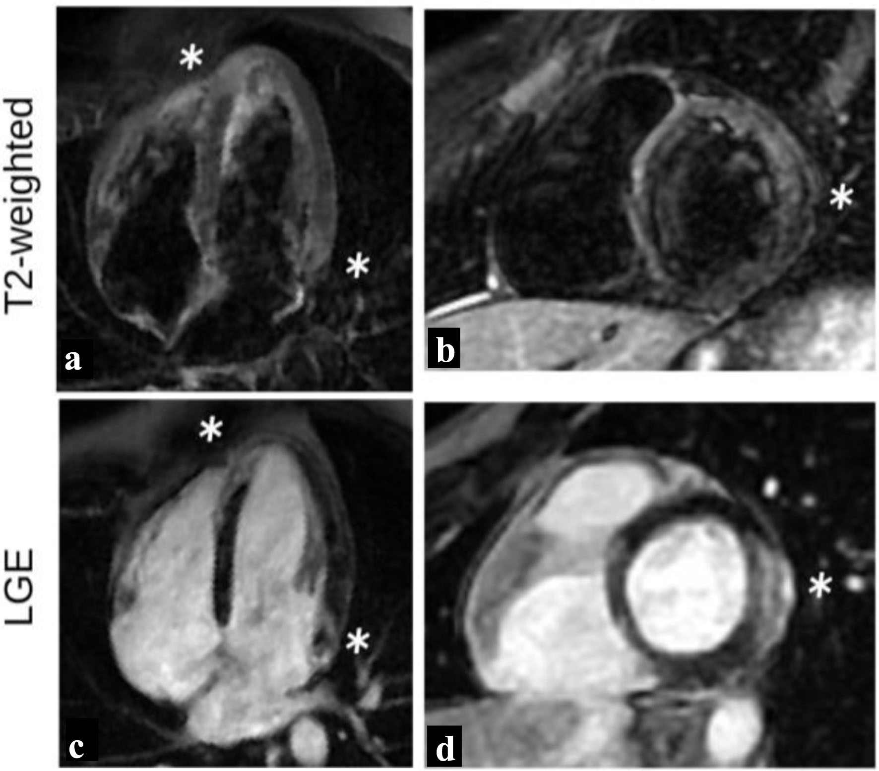
Figure 3. T2-weighted images and LGE distribution during initial study. High intensity signal (white stars) of the basal lateral and apical septum wall segments on T2-weighted images (a, b) indicating myocardial edema, with associated LGE of the same areas (c, d), findings indicative of acute myocarditis. LGE: late gadolinium enhancement.
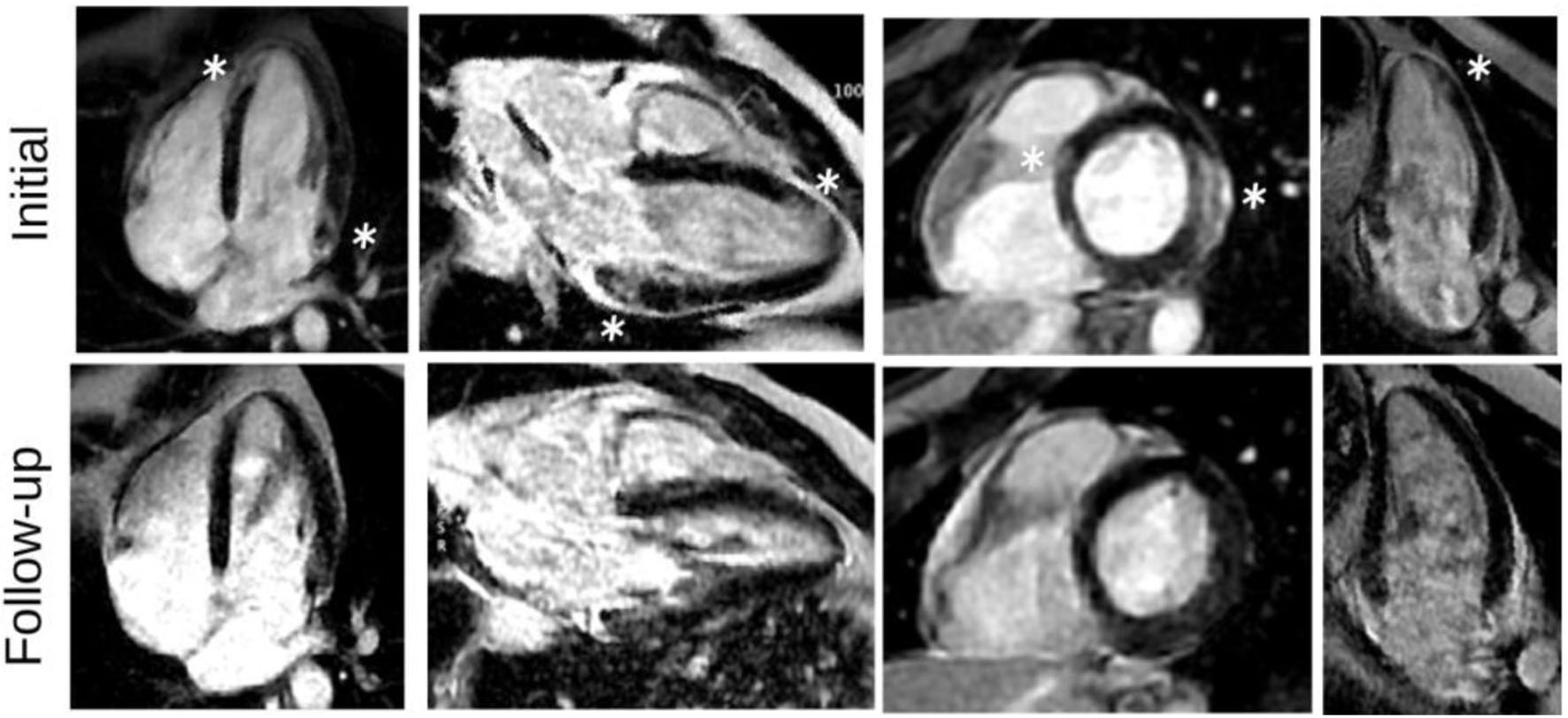
Figure 4. LGE images, during the initial presentation and in follow-up study. LGE images show subepicardial LGE of the basal lateral and apical septum wall segments (white stars), as well as patchy LGE distribution in a nonischemic pattern with significant improvement on follow-up. LGE: late gadolinium enhancement.