Journal of Medical Cases, ISSN 1923-4155 print, 1923-4163 online, Open Access
Article copyright, the authors; Journal compilation copyright, J Med Cases and Elmer Press Inc
Journal website https://www.journalmc.org

Case Report

Volume 14, Number 4, April 2023, pages 124-129


Rapidly-Progressing Pyomyositis After Chest Contusion in a Patient With Well-Controlled Diabetes Mellitus

Figures

Figure 1.
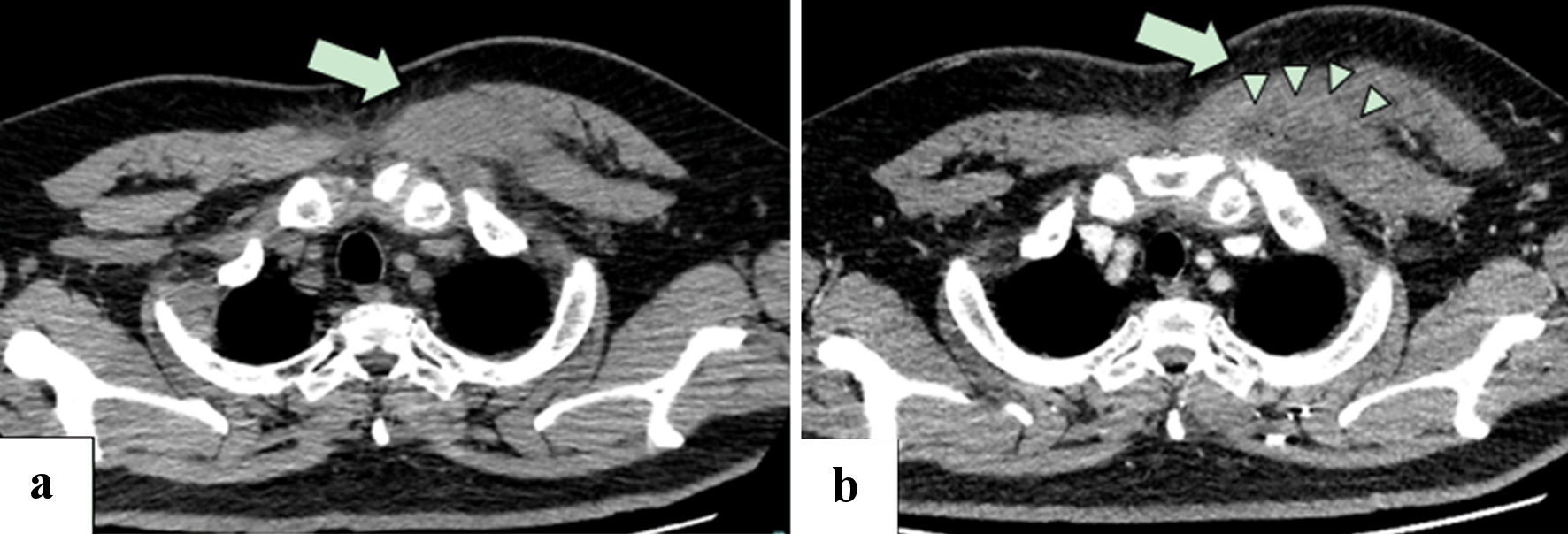
Figure 1. (a) Plain CT of the chest 1 day before admission showed swelling of the left major pectoralis muscle, with subcutaneous fat stranding (arrow). (b) Contrast-enhanced CT of the chest on day 3 of hospitalization showed a poor contrast enhancement area in the swelling of the major pectoralis muscle with ring-enhancement (arrow) and minute gas production (arrow heads), suggesting abscess formation. Left pleural effusion was revealed. CT: computed tomography.
Figure 2.
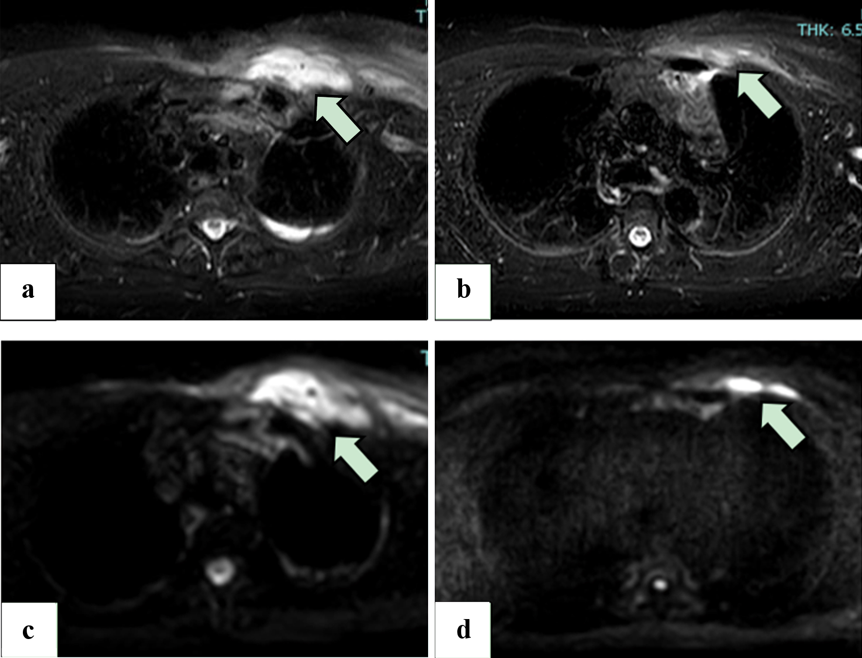
Figure 2. (a, b) STIR images from the MRI scan on day 10 of hospitalization (a) and 11 days after hospital discharge (b), without intravenous administration of gadolinium. Areas of high signal (arrow) reduction indicate that the swelling of the left pectoralis major has improved. (c, d) DWI MRI scans of the chest on day 10 of hospitalization (c) and 11 days after hospital discharge (d), without intravenous administration of gadolinium. The volume of the abscesses in the pectoralis major muscle (arrow) has been reduced. MRI: magnetic resonance imaging; STIR: short tau inversion recovery; DWI: diffusion-weighted imaging.

Tables

Table 1. Laboratory Data on Admission
 
WBC: white cell count; Ht: hematocrit; RBC: red cell count; Hb: hemoglobin; Plt: platelet count; TP: total protein; Alb: albumin; BUN: blood urea nitrogen; Cre: creatinine; T-Bil: total bilirubin; AST: aspartate aminotransferase; ALT: alanine aminotransferase; γGTP: gamma-glutamyl transpeptidase; ALP: alkaline phosphatase; LDH: lactate dehydrogenase; CK: creatine kinase; CRP: C-reactive protein; HbA1c: hemoglobin A1c; IgM: immunoglobulin M; IgG: immunoglobulin G; IgA: immunoglobulin A; HBs-Ag: hepatitis B surface antigens; HCV-Ab: hepatitis C antibody; COI: cut-off index.
Complete blood count
  WBC10,300/µL
  Neutrophils85.0%
  Lymphocytes11.0%
  Monocytes3.0%
  Eosinophils1.0%
  Basophils-
  Ht44.1%
  RBC515 × 104/µL
  Hb15.1 g/dL
  Plt14.2 × 104/µL
Biochemical examinations
  TP6.1 g/dL
  Alb3.7 g/dL
  BUN15.7 mg/dL
  Cre0.93 mg/dL
  T-Bil2.0 mg/dL
  AST136 U/L
  ALT138 U/L
  γGTP178 U/L
  ALP135 U/L
  LDH291 U/L
  CK198 U/L
CRP22.12 mg/dL
Glucose126 mg/dL
HbA1c6.7%
Glycoalbumin16.0%
Immunology and infection
  IgM89 mg/dL
  IgG709 mg/dL
  IgA180 mg/dL
  C3218 mg/dL
  C447 mg/dL
  CH5088.1 U/mL
  HBs-Ag0.00 IU/mL
  HCV-Ab0.01 COI


Table 2. Clinical Stages of Pyomyositis [8-10] and the Clinical Course of the Present Case
 
Disease stageClinical characteristicsManagementThe clinical course of the present case
aDay -5 indicates 5 days prior to the patient’s admission. bDay -3 indicates 3 days prior to the patient’s admission.
Stage 1 (invasive stage)Usually lasts 10 - 21 daysAntimicrobial therapyaDay -5: muscle blunt trauma
Swelling, pain
Low-grade fever, malaise
Often mistaken for contusion, hematoma, or muscle strain
Stage 2 (suppurative stage)Duration from 24 h to 12 daysAntimicrobial therapy and drainagebDay -3: high-grade fever, muscle pain
Fever, chillsDay 0 (admission): severe pain and tenderness, bacteremia
Prominent tenderness
Abscess formation
Most patients present at this stage
Stage 3 (late stage)High-grade feverAntimicrobial therapy and drainageDay 3: abscess formation
Severe pain
Bacteremia, sepsis