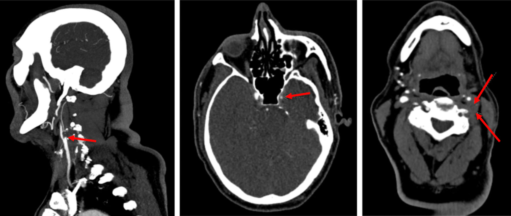
Figure 1. CTA of head and neck with red arrows in each image demonstrating complete occlusion of vertebral and internal carotid arteries. CTA: computed tomography angiography.
| Journal of Medical Cases, ISSN 1923-4155 print, 1923-4163 online, Open Access |
| Article copyright, the authors; Journal compilation copyright, J Med Cases and Elmer Press Inc |
| Journal website https://www.journalmc.org |
Case Report
Volume 14, Number 6, June 2023, pages 200-203
Identifying the Cause of Acute Left-Sided Visual Loss: A Clinical Dilemma
Figures