Journal of Medical Cases, ISSN 1923-4155 print, 1923-4163 online, Open Access
Article copyright, the authors; Journal compilation copyright, J Med Cases and Elmer Press Inc
Journal website https://www.journalmc.org

Case Report

Volume 14, Number 3, March 2023, pages 88-94


Extracranial Microvascular Complications of Moyamoya Disease Leading to Left Ventricular Systolic Dysfunction

Figures

Figure 1.
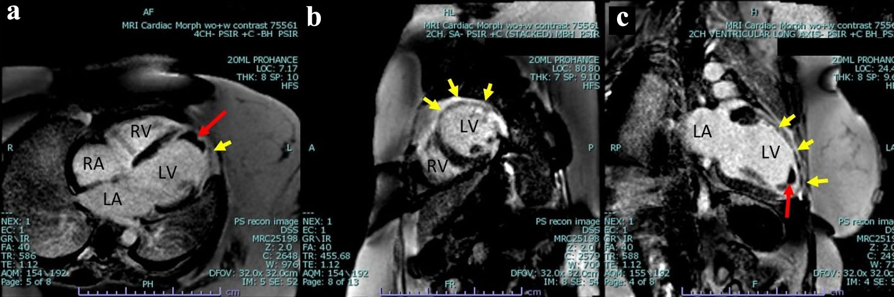
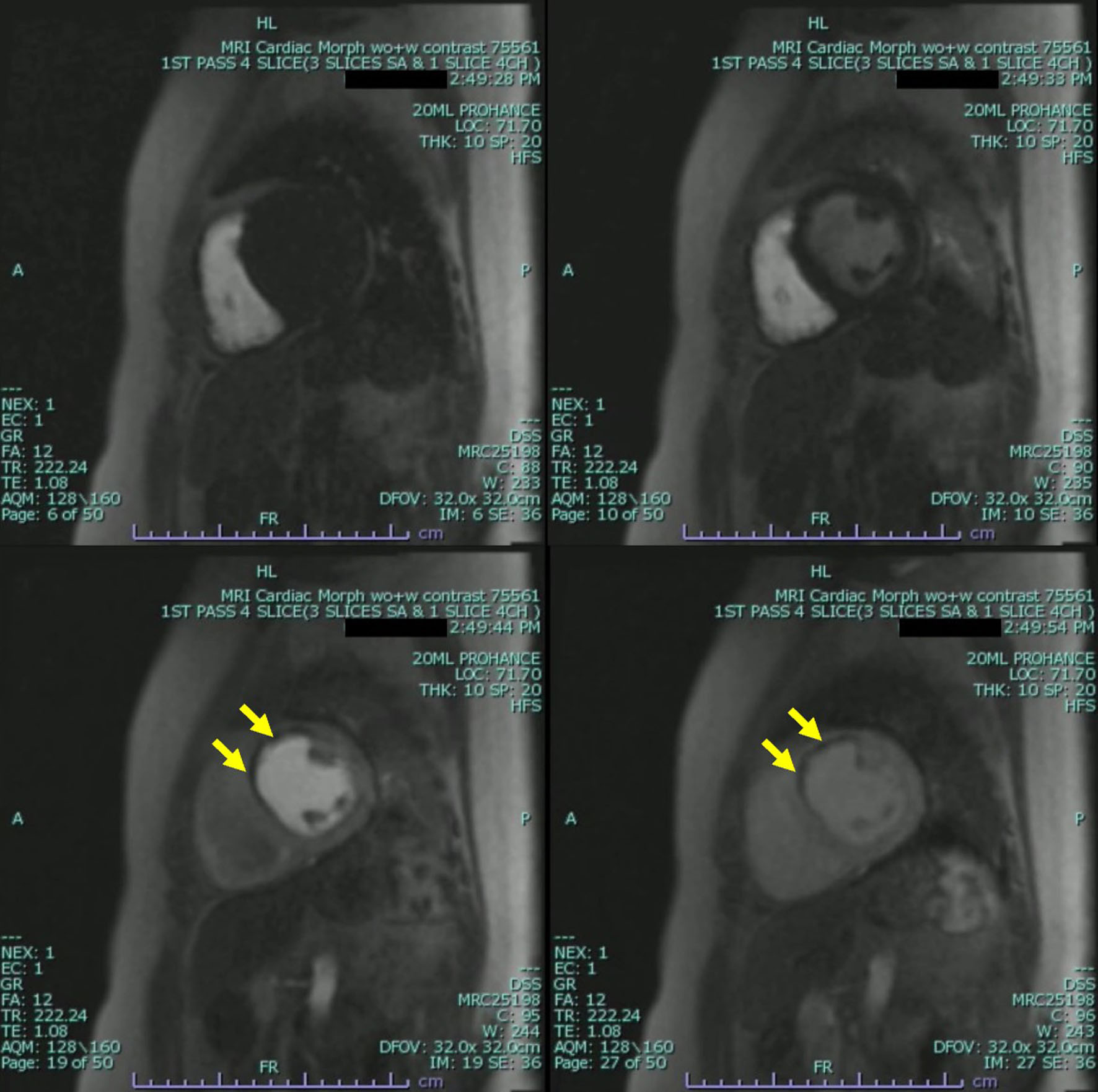
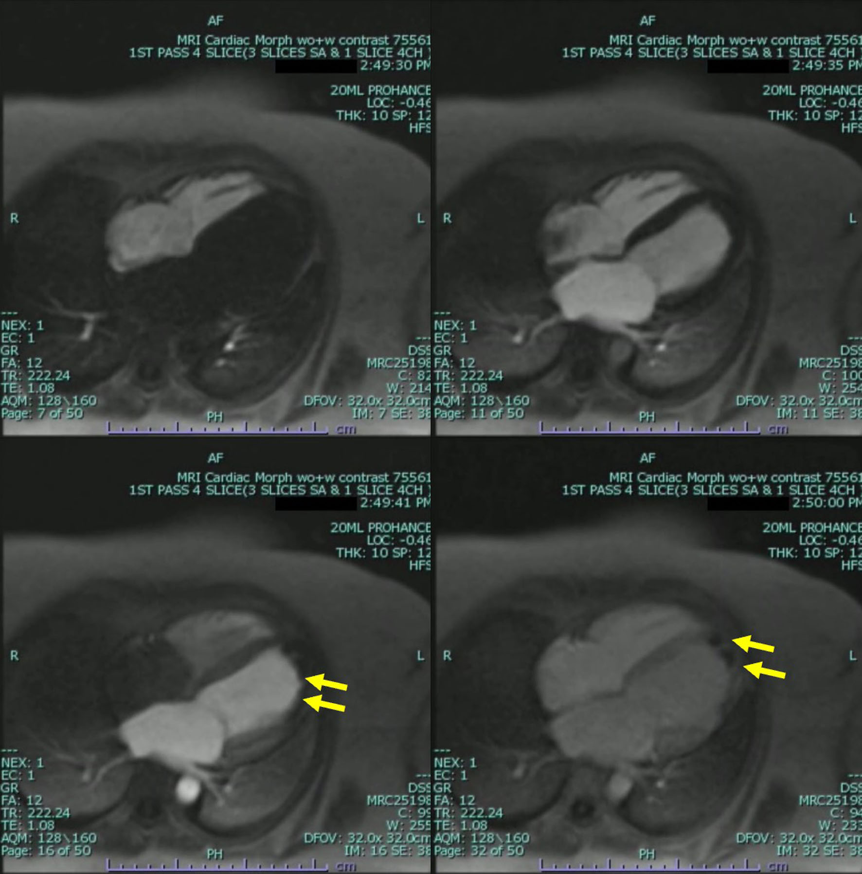
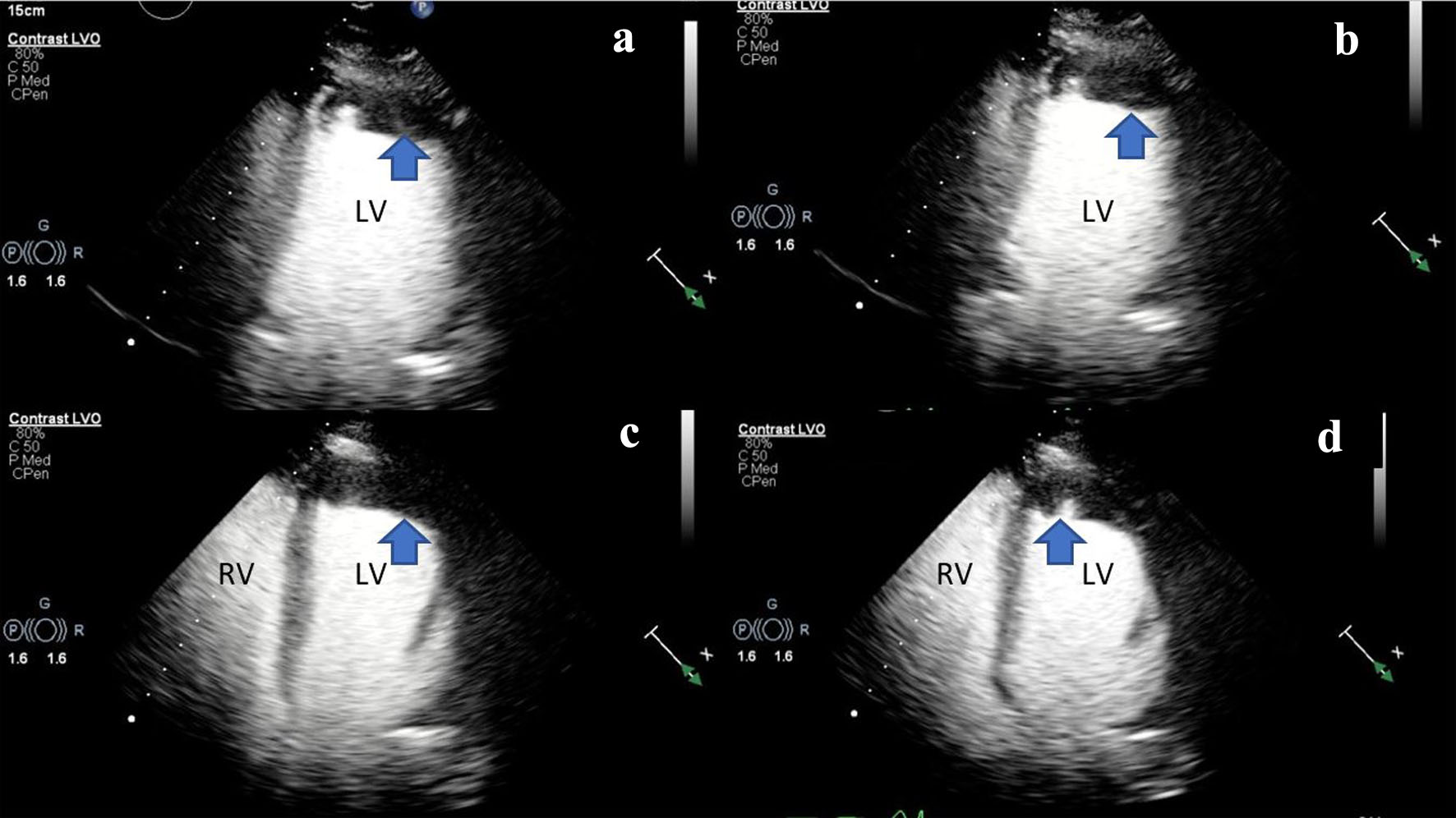
Figure 1. (a, b) Two-dimensional TTE (ultrasound enhancing agents) of an apical two-chamber view, at end-systole demonstrating severely reduced LV systolic function and LV apical hypokinesis with an associated LV apical thrombus (blue arrow). (c, d) Two-dimensional TTE still frame (ultrasound enhancing agents) of an apical four-chamber view, at mid-systole demonstrating biventricular dilation and an LV apical thrombus (blue arrow). Left ventricular internal diameter in diastole (LVIDd): 5.3 cm (3.7 - 5.5 cm), left ventricular internal diameter in systole (LVIDs): 4.7 cm (2.0 - 4.0 cm), right ventricular internal diameter in diastole (RVDd): 3.7 cm (1.9 - 3.5 cm). LV: left ventricle; RV: right ventricle; TTE: transthoracic echocardiogram.
Figure 2.
Figure 2. Cardiac magnetic resonance imaging phase-sensitive inversion recovery (CMRI PSIR) contrast-enhanced tissue always displays a higher signal (lighter) than normal myocardium (darker). (a) Four-chamber view, large thrombus measuring 3.0 × 1.2 cm in the apex and apical anterior wall (red arrow) as well as greater than 75% subendocardial late gadolinium enhancement in the mid anterior wall (yellow arrow). (b) Two-chamber short axis view demonstrating near transmural late gadolinium enhancement of the mid lateral wall transmural as well as greater than 75% subendocardial late gadolinium enhancement in the mid anterior wall (yellow arrow). (c) Two-chamber ventricular long axis view demonstrating a large thrombus measuring 3.0 × 1.2 cm in the apex and apical anterior wall (red arrow), as well as near transmural late gadolinium enhancement of the mid lateral wall transmural as well as greater than 75% subendocardial late gadolinium enhancement in the mid anterior wall and apex (yellow arrow). LV: left ventricle; RV: right ventricle; RA: right atrium; LA: left atrium.
Figure 3.
Figure 3. Cardiac magnetic resonance imaging (CMRI) two-chamber short axis first-pass perfusion imaging sequences still frame with injection of gadolinium demonstrating absence of uptake on first-pass perfusion of the mid anterior wall consistent with infarct pattern (yellow arrow).
Figure 4.
Figure 4. Cardiac magnetic resonance imaging (CMRI) two-chamber short axis first-pass perfusion imaging sequences still frames with injection of gadolinium demonstrating absence of uptake on first-pass perfusion of the apical and mid lateral wall consistent with infarct pattern. Apical mass is also demonstrated with absence of uptake on first-pass perfusion indicating a mass without native blood supply consistent with a thrombus (yellow arrow).

Table

Table 1. Cases of Moyamoya Disease Associated With Coronary Artery Disease, Adapted From Komiyama et al [14]
 
Case no.Author (years)Age at presentation of CADSexEthnicityCoronary manifestationsCoronary angiographyTreatment for CAD
CAD: coronary artery disease; ADA: anterior descending artery; CA: coronary artery; CxA: circumflex artery; MT: main trunk; lt: left; rt: right; bil: bilateral.
1Furuta et al (1985) [16]14MJapaneseNoneStenosis of rt CABypass surgery
2Shiratori et al (1985) [17]29FJapaneseAngina pectorisStenosis at ostium of lt CA MT, irregular wall at bil CAsBypass surgery
3Sakamoto et al (1987) [18]31MJapaneseAngina pectorisStenoses of rt CA and lt ADABypass surgery
4Saito et al (1987) [19]36MJapaneseAngina pectorisStenosis of lt ADAUnavailable
5Tateno et al (1988) [20]30FJapaneseAngina pectorisStenosis of lt ADAUnavailable
6Suzuki et al (1989) [21]40FJapaneseHeart failureStenosis at ostium of lt CA MT, atherosclerosis of lt ADAUnavailable
7Tokunaga et al (1996) [22]34MJapaneseSilent myocardial ischemiaHypoplastic lt CxA, diffuse scleroses of rt CA and lt ADANone
8Akasaki et al (1998) [23]23MJapaneseAngina pectorisStenosis of lt ADANone
9Ikeda et al (1998) [24]48MJapaneseAngina pectorisNormal, positive to Ach provocationUnavailable
10Ahn et al (1999) [25]19FKoreanMyocardial infarctionStenosis of middle lt ADANone
11Goar et al (1999) [26]36MIndianAngina pectorisStenoses at ostium of lt CA MT and rt CANone