Journal of Medical Cases, ISSN 1923-4155 print, 1923-4163 online, Open Access
Article copyright, the authors; Journal compilation copyright, J Med Cases and Elmer Press Inc
Journal website https://www.journalmc.org

Case Report

Volume 13, Number 11, November 2022, pages 536-540


A Rare and Unusual Cause of Ischemic Stroke to Be Aware of

Figures

Figure 1.
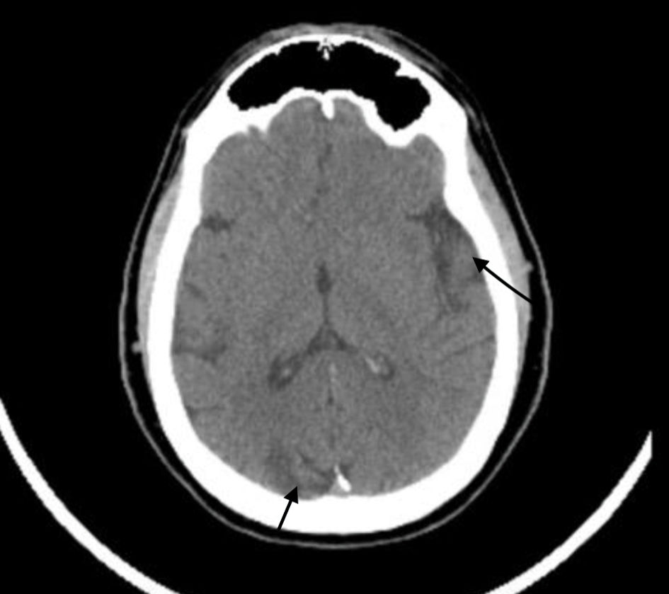
Figure 1. Computed tomography of the head without contrast showing multifocal areas of encephalomalacia and gliosis within the left parietal lobe (long black arrow on right), right occipital lobe (short black arrow at bottom) and left cerebellum. No signs of hemorrhage were noted.
Figure 2.
Figure 2. Computed tomography angiogram of the head and neck showing left external (yellow arrow) and internal carotid artery (white arrow) with an isolated right external (yellow arrow) carotid artery with the absence of right internal carotid branch.