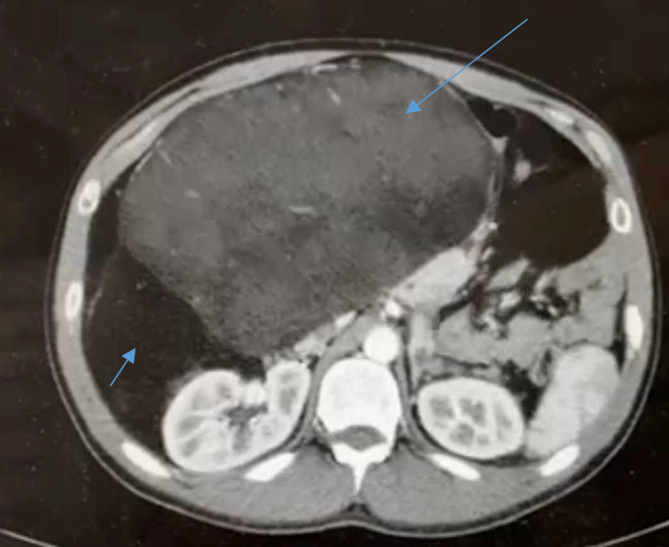
Figure 1. An axial abdominopelvic CT scan demonstrates a huge retroperitoneal mass with area of high (long arrow) and low (short arrow) of density, and the mass displacing the abdominal organs to the left side. CT: computed tomography.
| Journal of Medical Cases, ISSN 1923-4155 print, 1923-4163 online, Open Access |
| Article copyright, the authors; Journal compilation copyright, J Med Cases and Elmer Press Inc |
| Journal website https://www.journalmc.org |
Case Report
Volume 13, Number 10, October 2022, pages 517-520
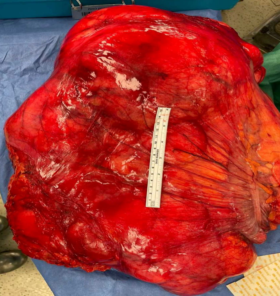
Retroperitoneal Liposarcoma: The Giant Type
Figures