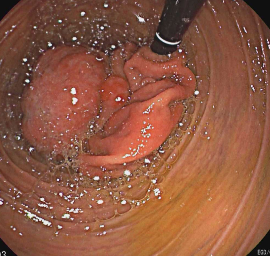
Figure 1. Endoscopy showing duodenum with remnant of stomach on retroflexion.
| Journal of Medical Cases, ISSN 1923-4155 print, 1923-4163 online, Open Access |
| Article copyright, the authors; Journal compilation copyright, J Med Cases and Elmer Press Inc |
| Journal website https://www.journalmc.org |
Case Report
Volume 13, Number 6, June 2022, pages 269-273
Cachexia and Invisible Stomach on Endoscopy: An Endoscopist’s Enigma and a Surgeon’s Axiom
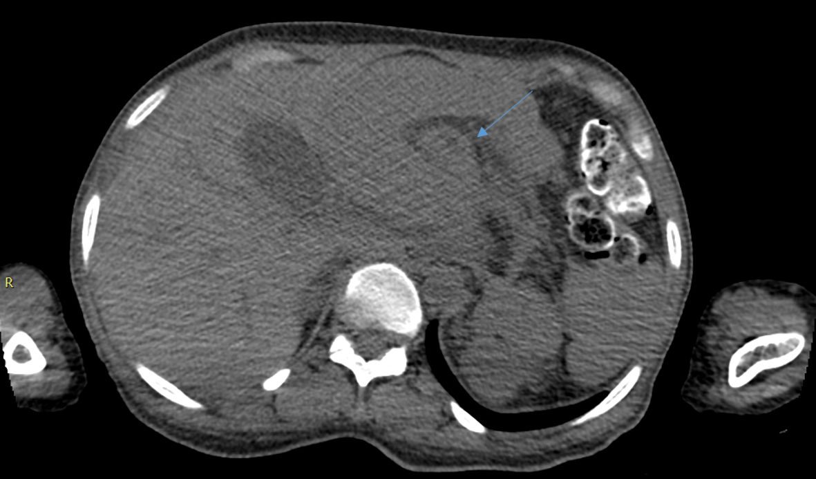
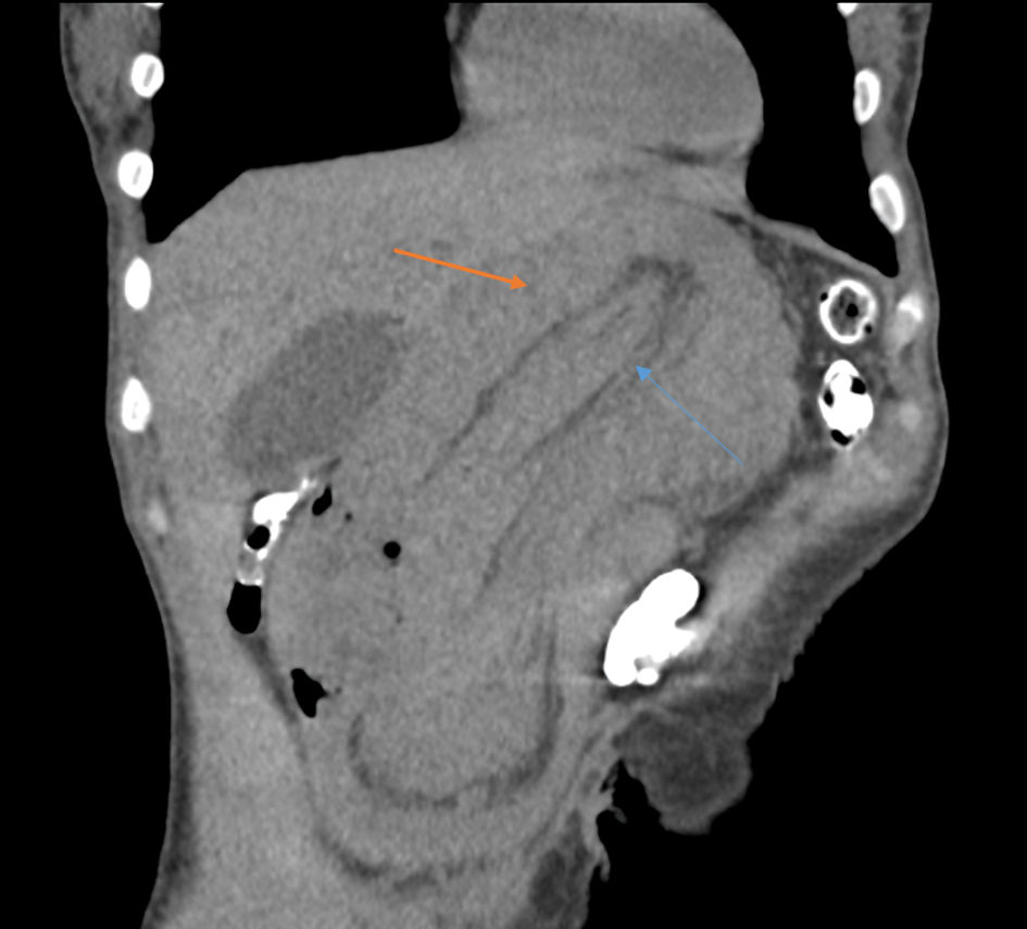
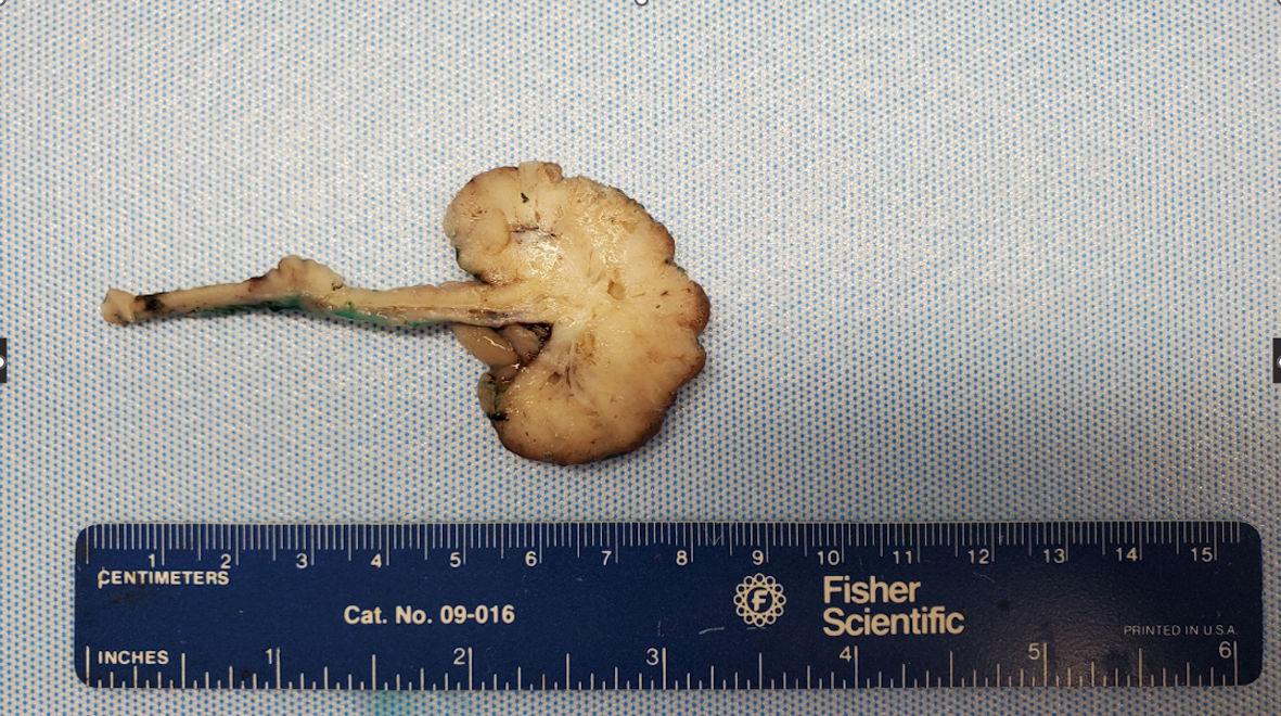
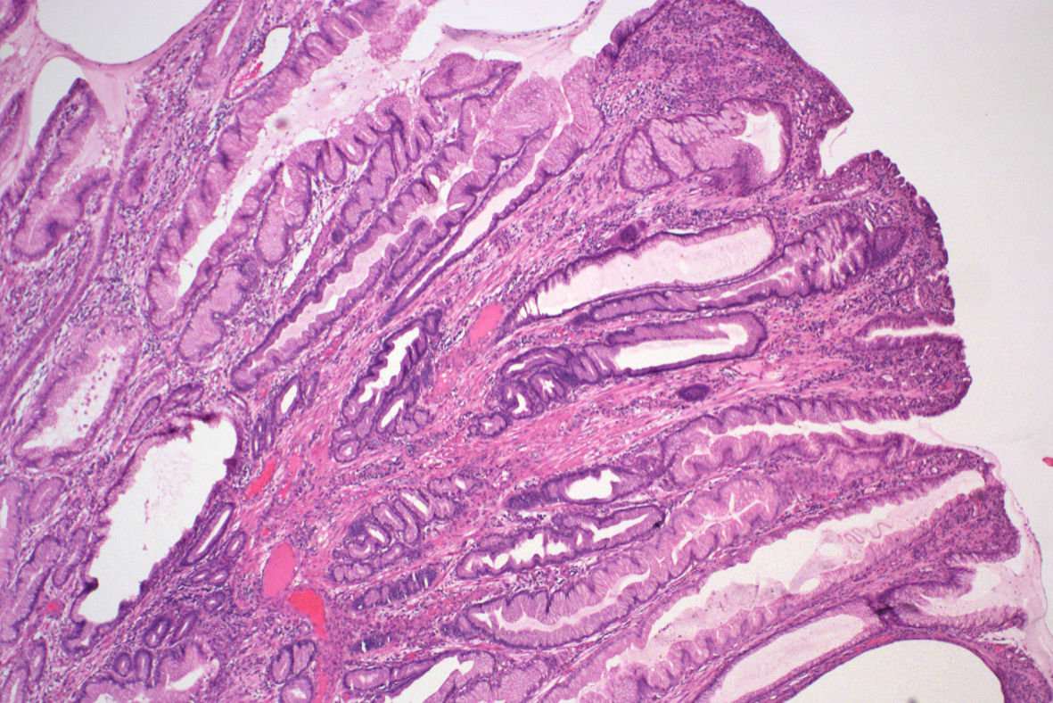
Figures