Journal of Medical Cases, ISSN 1923-4155 print, 1923-4163 online, Open Access
Article copyright, the authors; Journal compilation copyright, J Med Cases and Elmer Press Inc
Journal website https://www.journalmc.org

Case Report

Volume 13, Number 3, March 2022, pages 129-134


Immunoglobulin G4-Associated Cholangitis: Two Case Reports and Clinical Use of Classic and Emerging International Diagnostic Criteria

Figures

Figure 1.
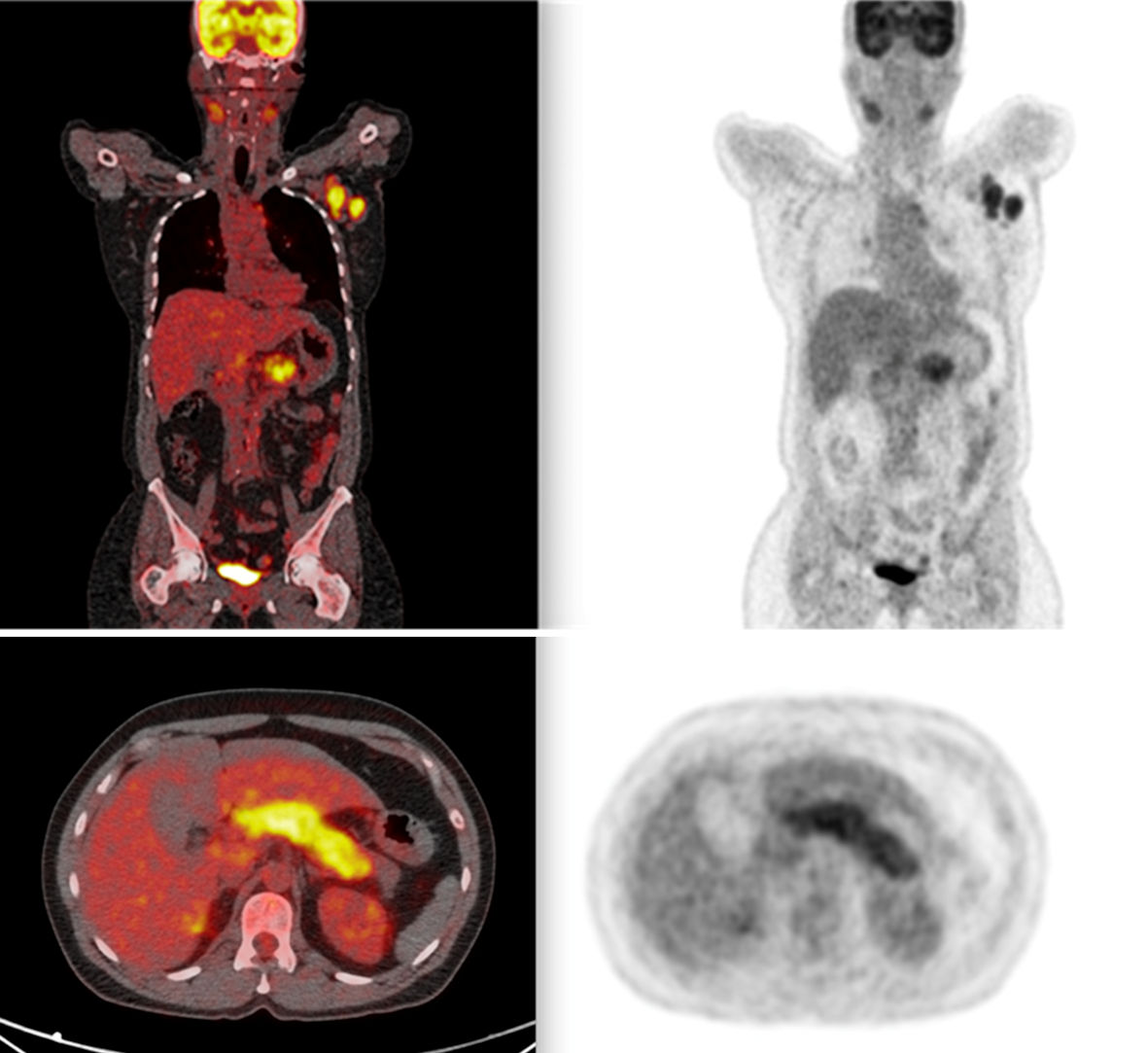
Figure 1. PET scan showing widespread lymph node involvement (pancreatic, submandibular, and hepatic hilum). In the pancreas, in addition to pancreatic enlargement, note the increased FDG uptake. PET: positron emission tomography; FDG: fluorodeoxyglucose.
Figure 2.
Figure 2. Narrowing of the left biliary tree with retraction of the hepatic parenchyma.
Figure 3.
Figure 3. Fluoroscopy showing compression at the confluence of the hepatic ducts.
Figure 4.
Figure 4. Review of original biopsy, with evidence of storiform fibrosis on hematoxylin and eosin staining and IgG4+ plasma cells on immunohistochemistry. Ig: immunoglobulin.

Tables

Table 1. Laboratory Findings of Case 1
 
TestAdmissionFollow-up
GGT: gamma-glutamyl transferase.
Alkaline phosphatase400 U/L192 U/L
GGT200 U/L79 U/L
Total bilirubin2.0 mg/dL1.2 mg/dL


Table 2. Laboratory Findings of Case 2
 
TestAdmissionFollow-up
GGT: gamma-glutamyl transferase.
Alkaline phosphatase1,080 U/L800 U/L
GGT104 U/L70 U/L
Total bilirubin10 mg/dL1.28 mg/dL