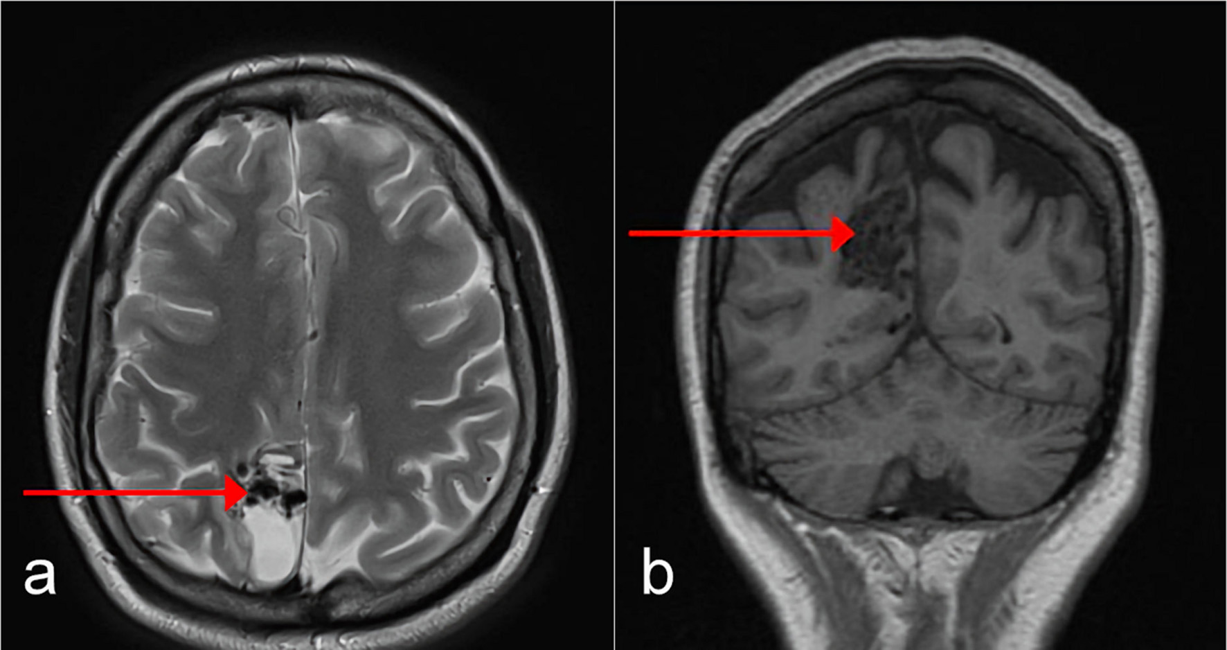
Figure 1. Axial (a) and coronal (b) MRI of the brain performed at our institution demonstrated a nidal type right parietal AVM (as shown by arrows). MRI: magnetic resonance imaging; AVM: arteriovenous malformation.
| Journal of Medical Cases, ISSN 1923-4155 print, 1923-4163 online, Open Access |
| Article copyright, the authors; Journal compilation copyright, J Med Cases and Elmer Press Inc |
| Journal website https://www.journalmc.org |
Case Report
Volume 13, Number 3, March 2022, pages 104-108
An Unusual Case of Cerebral Arteriovenous Malformation in Pregnancy
Figures