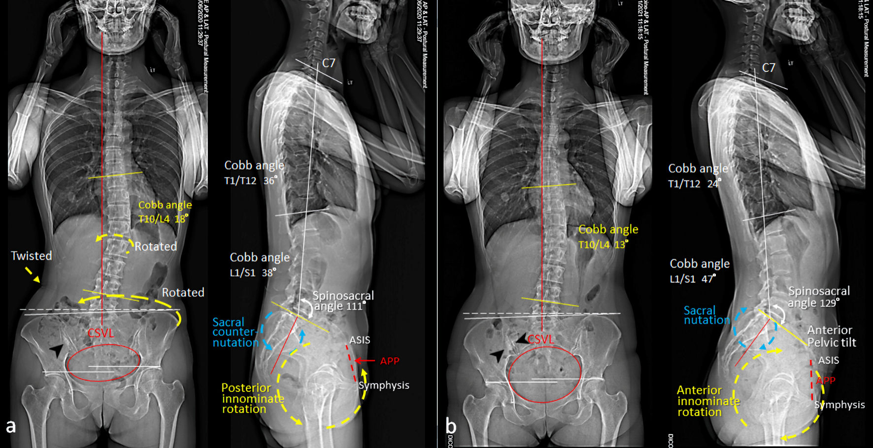
Figure 1. Comparison of standing assessment pre- and post-treatment. (a) Initial radiographs revealed an inflammatory sclerosis (black arrow head) on the iliac side of the right sacroiliac joint (SIJ), sacral counter-nutation (backward rotation, blue circle), posterior pelvic tilt (yellow circle), forward left innominate and twisted lumbar spine. (b) Six months later, radiographs showed dense sclerosis surrounding the right SIJ (black arrow heads), sacral nutation (forward rotation, blue circle) and anterior pelvic tilt (yellow circle). Normally, the range of motion of the SIJ does not exceed 3° and nutation occurs during increased load-bearing situations e.g., standing and sitting. APP: anterior pelvic plane (dashed red line); ASIS: anterior superior iliac spine; CSVL: central sacral vertical line (red line).
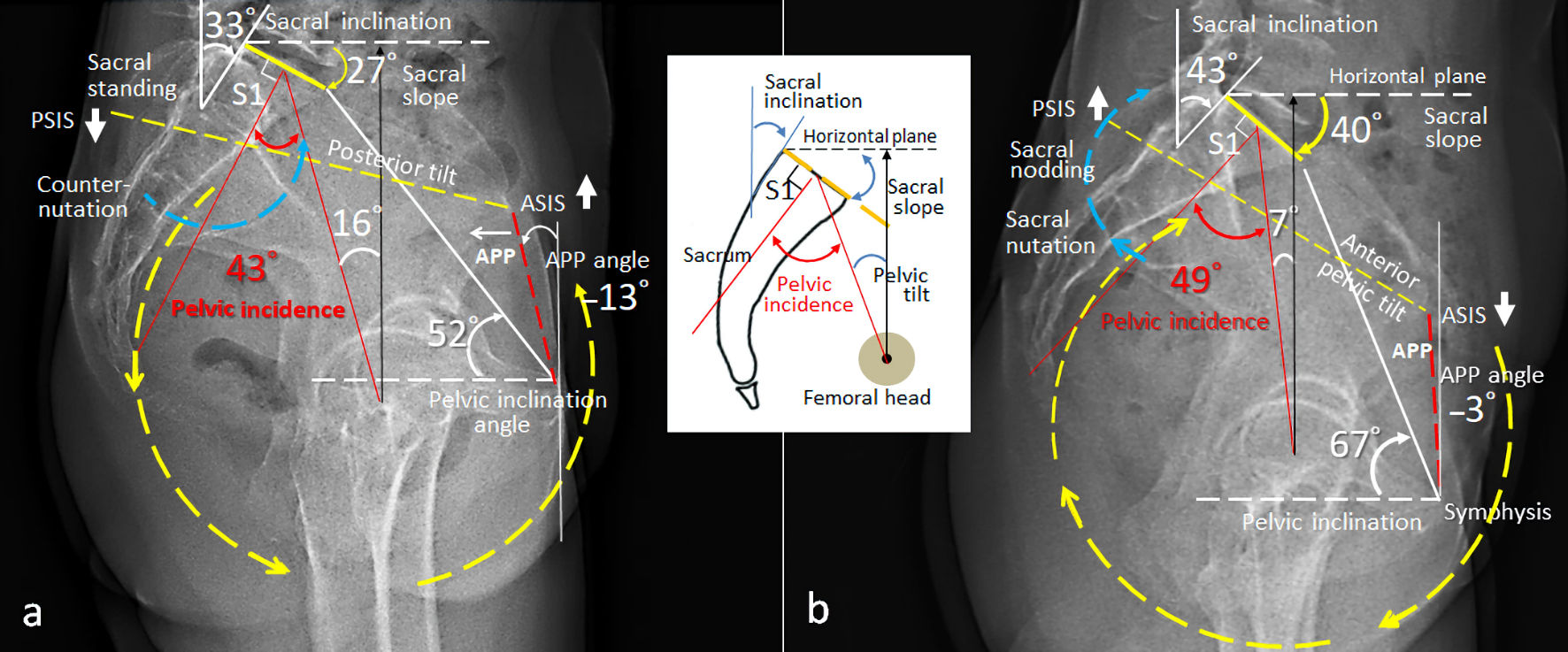
Figure 2. Comparison of spinopelvic parameters measured before and after chiropractic care. (a) Pre-treatment radiograph revealed counter-nutation of the sacrum (blue circle), sacral inclination 33°, sacral slope 27°, posterior innominate rotation (yellow circle), and posterior pelvic tilt (APP angle -13°, pelvic inclination 52°). (b) Six months later, interval change in all the measurements was demonstrated on the follow-up radiograph. Pelvic incidence (PI) has been considered as a static parameter in health individuals. Significant PI change (43° vs. 49°) between two consecutive radiographs is suggestive of sacroiliac instability. APP: anterior pelvic plane (dashed red line); ASIS: anterior superior iliac spine; PSIS: posterior superior iliac spine. A schematic depicting geometries for the pelvic measurements is inserted.