Journal of Medical Cases, ISSN 1923-4155 print, 1923-4163 online, Open Access
Article copyright, the authors; Journal compilation copyright, J Med Cases and Elmer Press Inc
Journal website https://www.journalmc.org

Case Report

Volume 12, Number 12, December 2021, pages 503-510


Male Adenoid Cystic Carcinoma of the Breast

Figures

Figure 1.
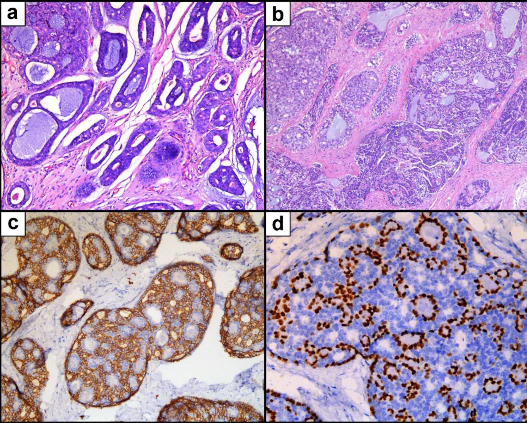
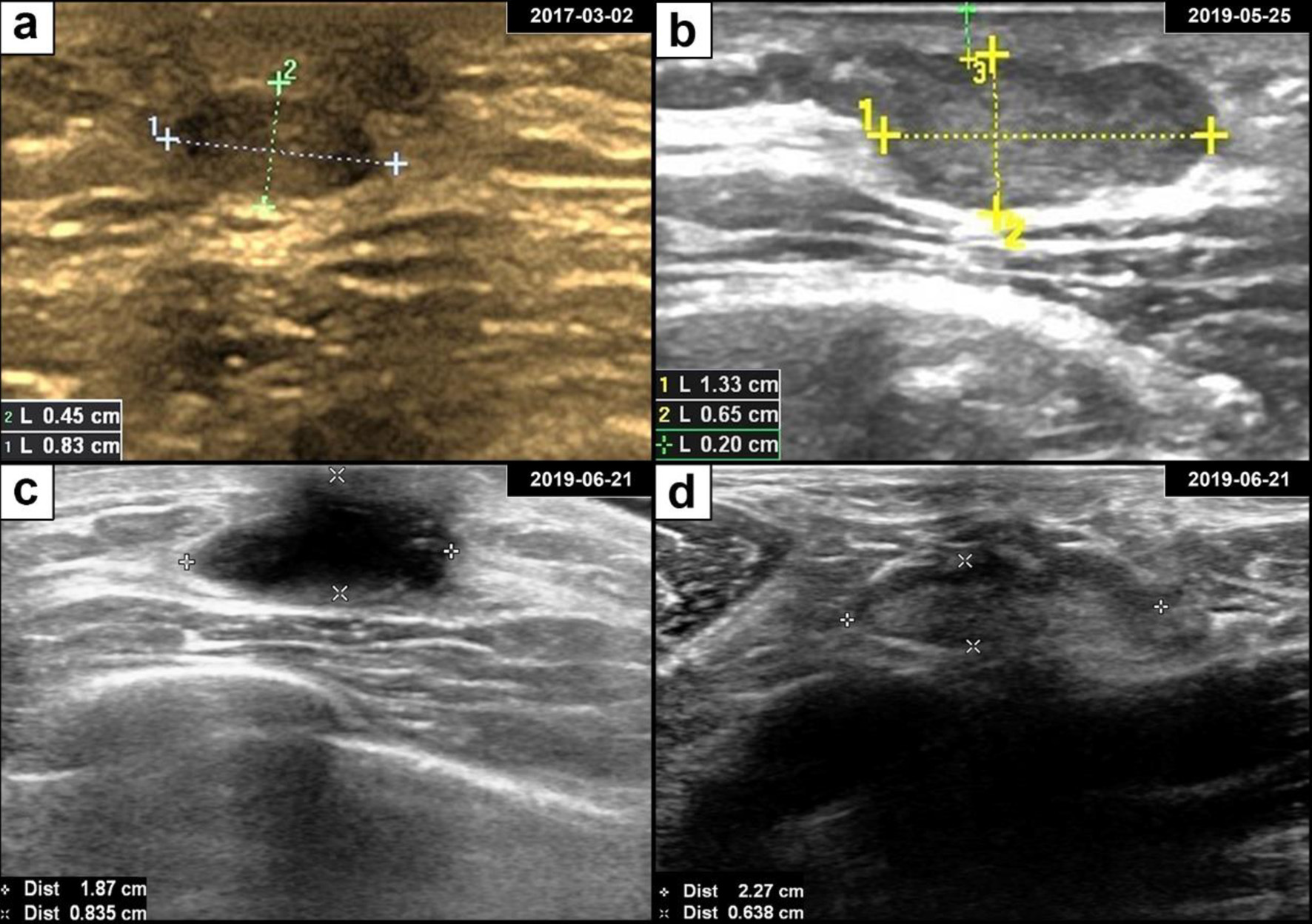
Figure 1. Breast adenoid cystic carcinoma ultrasound and postoperative findings. (a) Ultrasonography revealed a well-defined oval hypoechoic mass in the retroareolar region of the left breast. (b) The subareolar mass progressively enlarged over the course of 2 years. (c) Ultrasonography showed a hypoechoic area after lumpectomy. (d) Ultrasonography of an enlarged axillary lymph node with a clear boundary.
Figure 2.
Figure 2. Microscopic pathology of breast adenoid cystic carcinoma. (a) H&E staining showing an cribriform growth pattern. (b) H&E staining showing tubular and solid growth patterns. (c) IHC staining for CD117. (d) IHC staining for P63. H&E: hematoxylin and eosin stain; IHC: immunohistochemistry.
Figure 3.
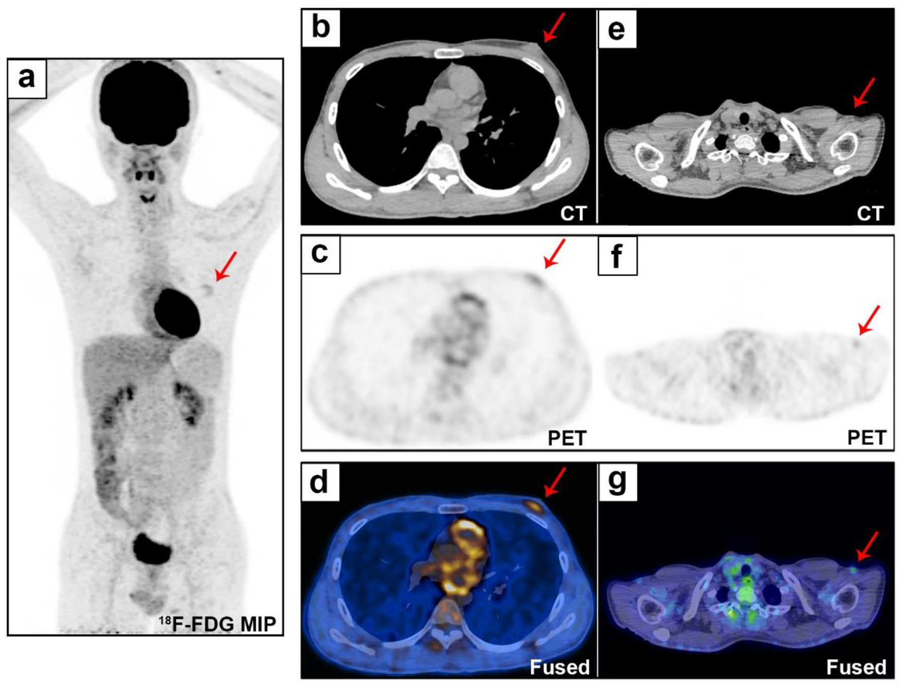
Figure 3. Whole-body evaluation by 18F-FDG PET/CT. The 18F-FDG PET/CT MIP (a) shows uptake (SUVmax 2.5) in the left breast lesion (b and d, red arrows), with a disordered tissue structure (c, red arrow). Small lymph nodes were found in the bilateral neck and bilateral axillary regions (e-g, red arrows, SUVmax 1.65). 18F-FDG PET/CT: 18F-fluorodeoxyglucose positron emission tomography/computed tomography; MIP: maximum intensity projection; SUVmax: maximum standardized uptake value.

Table

Table 1. Summary of Male Adenoid Cystic Carcinoma of Breast
 
ReferencesYearAgeDiameterNodeSurgeryAdjuvant treatmentOutcome of follow up
Neg: negative; Pos: positive; NR: no report; LM: lumpectomy; MRM: modified radical mastectomy; ALND: axillary lymph node dissection; ScM: subcutaneous mastectomy; SM: simple mastectomy; SNB: sentinel node biopsy; RM: radical mastectomy.
Presenting case2021231.3 cmNegLM → MRM + ALNDNoneRemain well after 24 months of follow-up
Hogan and Fan [20]2020602.1 cmNRLMRadiotherapyLung metastasis
Pang et al [19]2019441.2 cmNegLM → MRM + ALNDPostoperative chemotherapyRemain well after 26 months of follow-up
Douglas and Mackenzie [18]2019215.0 cmNegScM → SM + SNBNoneRemain well after 12 months of follow-up
Zadeh et al [17]2017424.0 cmNegMRM + ALNDNoneRemain well after 12 months of follow-up
Tang et al [16]2015193.0 cmNegRM + ALNDEndocrine therapyRemain well after 67 months of follow-up
Yoo et al [15]2013411.7 cmPosNRNoneBone and lung metastasis
Sahan et al [14]2012601.3 cmNRLM → RMNoneNR
Liu et al [13]2012202.1 cmNegSM + SNBNoneNR
Kshirsagar et al [11]200682NR3/5 PosMRM + ALNDRadiotherapyRecurrence at 2 years
Maciag et al [12]2006804 cm1/10 PosLM → SM + ALNDRadiotherapy + endocrine therapyRemain well after 5 years of follow-up
Miliauskas et al [10]1991133.8 cmNegLM → ScMNoneRemain well after 30 months of follow-up
Hjorth et al [9]197721NRNegSMNoneRemain well after 2 years of follow-up
Ferlito et al [8]197460NRNegSMNoneNR
Verani et al [7]1973783.5 cmNegMRM + ALNDNoneLung metastasis
Woyke et al [6]197037NRNegLMNoneRecurrence at 5 and 7 years
Ferlito [5]1969Middle agedNRNRNRNRNR