Journal of Medical Cases, ISSN 1923-4155 print, 1923-4163 online, Open Access
Article copyright, the authors; Journal compilation copyright, J Med Cases and Elmer Press Inc
Journal website https://www.journalmc.org

Case Report

Volume 12, Number 10, October 2021, pages 395-399


Stewart-Treves Syndrome in Obesity-Associated Chronic Lymphedema: A Case Report

Figures

Figure 1.
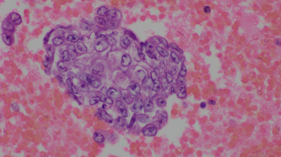
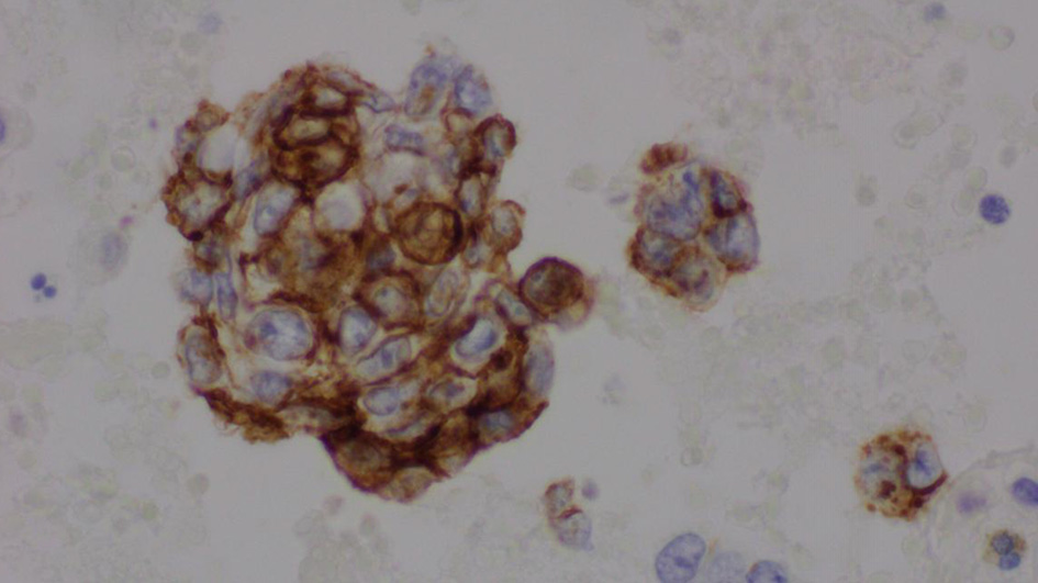
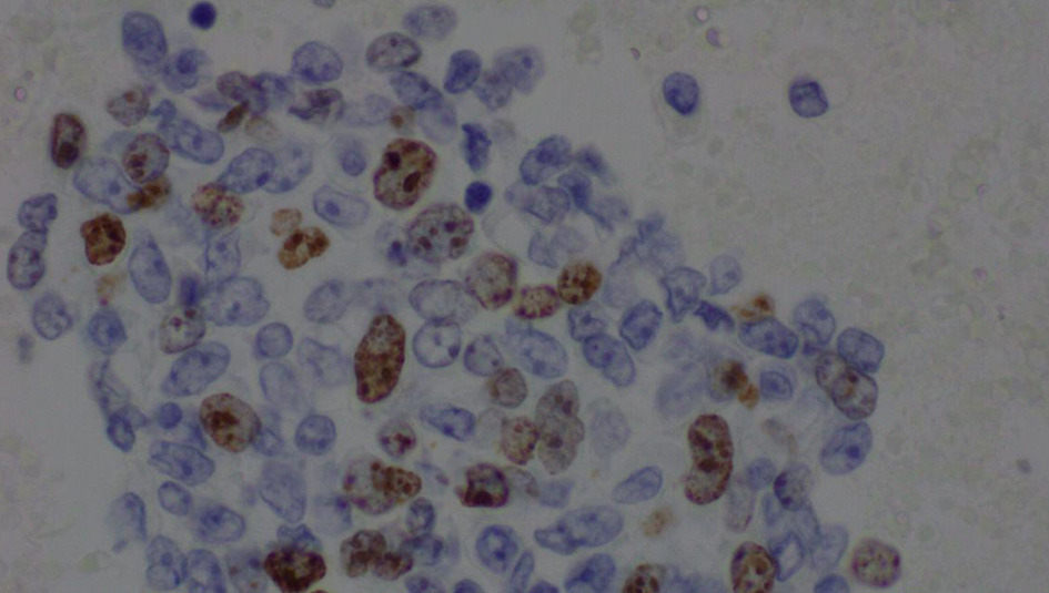
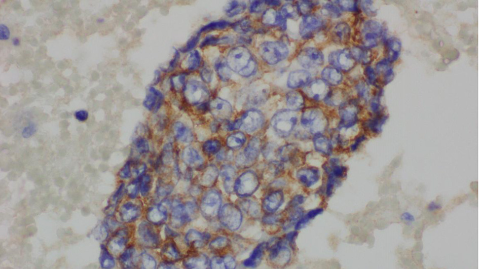
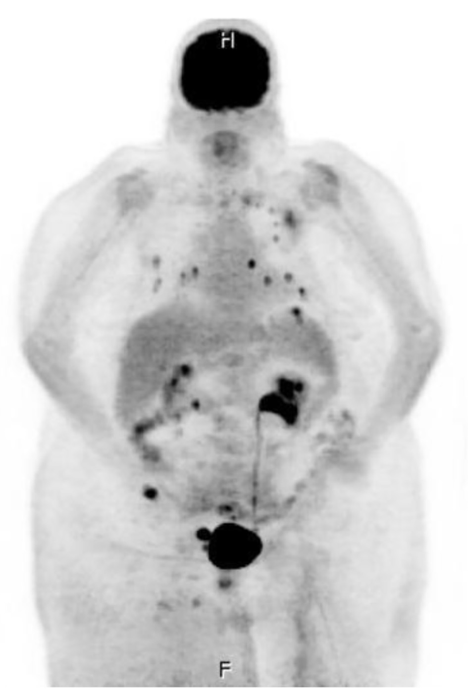
Figure 1. Whole-body positron emission tomography and computer tomography imaging were performed with multi-planar imaging without oral or intravenous contrast material, revealing metastatic disease to lymph node (right external iliac lymph node measuring 2.5 × 4.2 cm), bone (medial aspect of the left clavicle, the posterior lateral aspect of the head of the left humerus, the posterior aspect of the T2 vertebral body, and the right iliac wing), lung (there are approximately 15 solid noncalcified pulmonary nodules in each lung, ranging between 2 and 10 mm), and bilateral adrenal glands.
Figure 2.
Figure 2. Pleural fluid, cell block, H&E stain (× 600).
Figure 3.
Figure 3. Pleural fluid, cell block, immunohistochemical stain for CD34 (× 600).
Figure 4.
Figure 4. Pleural fluid, cell block, immunohistochemical stain for Ki-67 (× 600).
Figure 5.
Figure 5. Pleural fluid, immunohistochemical for epithelial membrane antigen (× 600).

Table

Table 1. Laboratory Values During Admission
 
Day 1Day 2Day 3Day 4Day 5
WBC: white blood cell; NV: normal value; BUN: blood urea nitrogen.
WBC (NV 4.1 - 11.0 × 10³/µL)16.512.716.821.119.2
Neutrophils (NV 50-76%)77.975.478.784.489.1
Hemoglobin (NV 12.0 - 16.0 g/dL)9.49.59.510.69.1
Platelet (NV 150 - 400 × 103/µL)357483515588506
BUN (NV 8 - 24 mg/dL)1717171823
Creatinine (NV 0.55 - 1.30 mg/dL)0.560.530.520.771.13
Lactic acid (NV 0.7 - 1.9 mmol/L)2.12.2