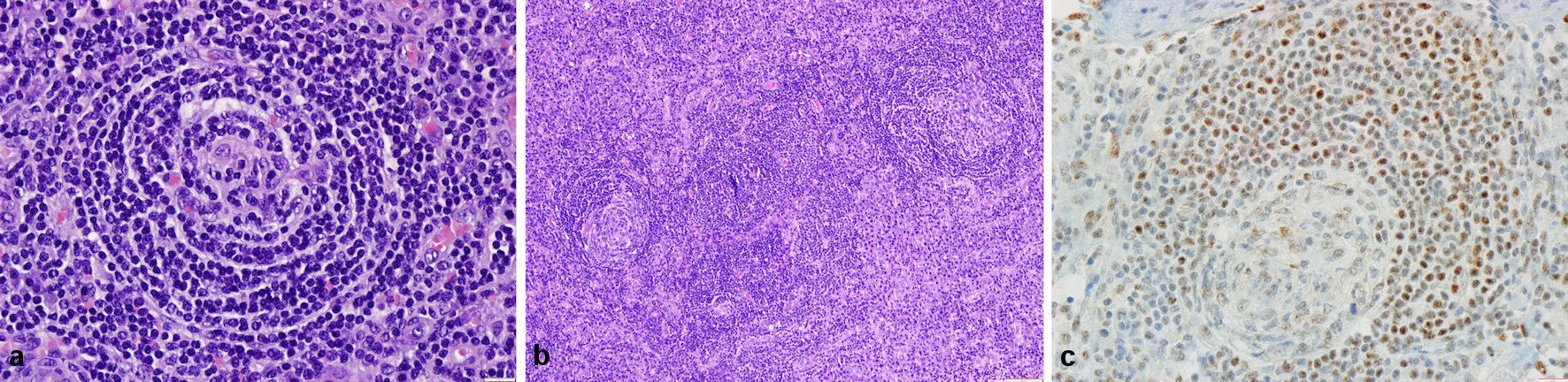
Figure 1. Histological findings in the surgical specimen. (a) A germinal center with lymphocyte depletion; small dark lymphocytes concentrically arranged in the mantle zone (hematoxylin and eosin (H&E), × 400). (b) Interfollicular regions expanded by sheets of plasma cells and vascular proliferation (H&E, × 100). (c) Immunohistochemical staining for human herpes virus-8 (HHV-8, × 400).
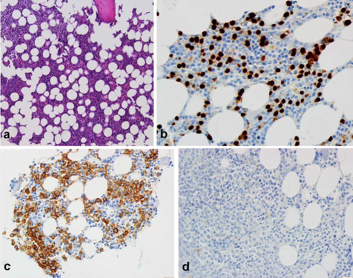
Figure 2. Histological findings in the bone marrow biopsy specimen. (a) Elements from the three hematopoietic lineages with normal density, location and maturation (hematoxylin and eosin (H&E), × 100). (b, c) Positive immunohistochemical staining for plasma cells accounting for about 5% of the total cell population ((b) CD138, × 400); (c) MUM1, × 400). (d) Negative immunohistochemical staining for human herpes virus-8 (HHV-8, × 400).