Journal of Medical Cases, ISSN 1923-4155 print, 1923-4163 online, Open Access
Article copyright, the authors; Journal compilation copyright, J Med Cases and Elmer Press Inc
Journal website https://www.journalmc.org

Case Report

Volume 12, Number 9, September 2021, pages 347-350


Unique Communication: A Case Report of Acquired Esophageal Pulmonary Fistula

Figures

Figure 1.
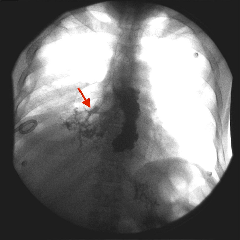
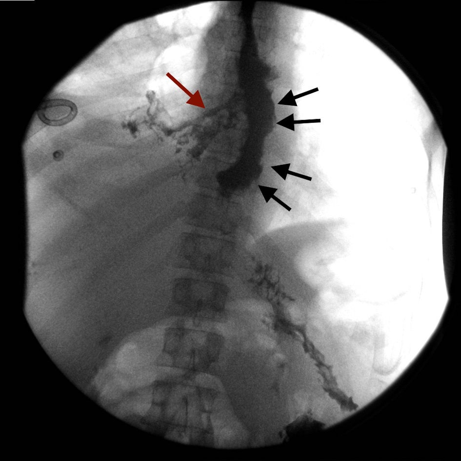
Figure 1. The distal one-third of esophagus is markedly dilated and irregular in contour (black arrows). Contrast is seen extravasating from the right side of the esophagus into the right lower lobe parenchyma consistent with esophageal pulmonary fistula (red arrow).
Figure 2.
Figure 2. Contrast is seen filling the right lower lobe bronchus, which suggests possible reflux from the right lower lung parenchyma (red arrow).