Figures
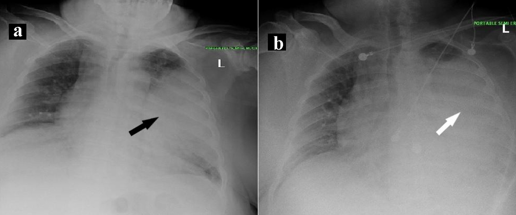
Figure 1. Chest X-ray findings during initial hospital admission. (a) Chest X-ray on day 1 of admission. Black arrow shows consolidation of left mid to lower lung field with possibility of an associated underlying mass. (b) Chest X-ray on day 3 of admission. White arrow shows near-complete opacification of the left hemithorax with minimal sparing of the apical region.
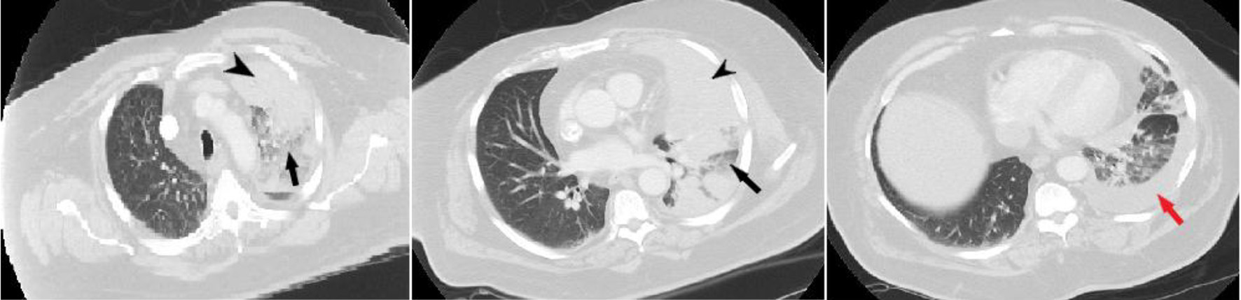
Figure 2. Results of chest computed tomography with contrast. Black arrowhead shows left upper lobe mass extending from chest wall to the mediastinum. Black arrow shows consolidation with air bronchograms. Red arrow shows left pleural effusion.
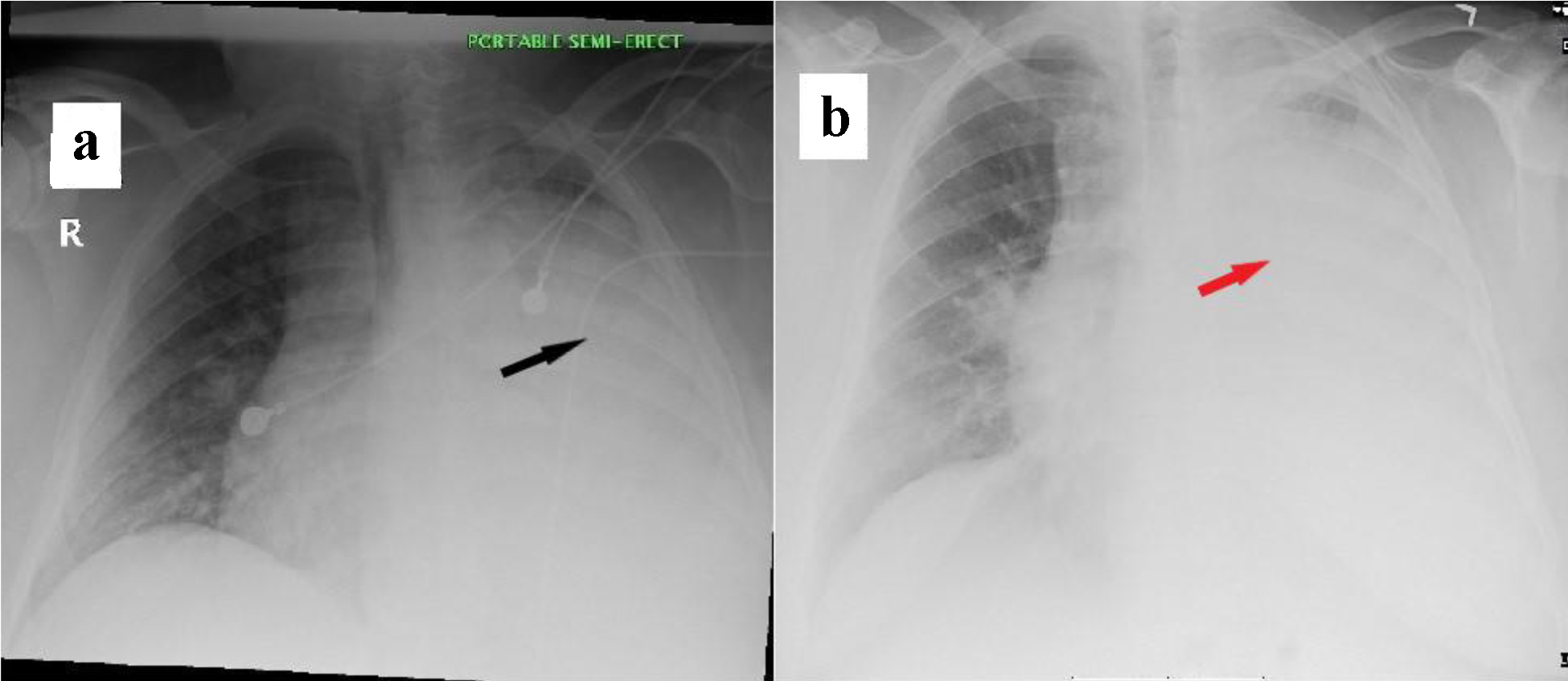
Figure 3. Chest X-ray findings during readmissions. (a) Chest X-ray on day of first readmission. Black arrow shows diffuse dense opacification of the left hemithorax with minimal sparing of the extreme apex. (b) Chest X-ray on day of second readmission. Red arrow shows opacification of left hemithorax with minimal sparing of the apex.
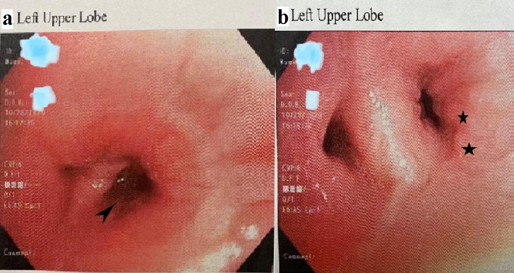
Figure 4. Images of bronchoscopy showing severe airway narrowing of left bronchi. (a) Left upper lobe (apical posterior). Arrowhead shows narrowing of the airway. (b) Left upper lobe (carina and lingula). Black star shows diffuse polypoid mucosal findings with irregular contours.
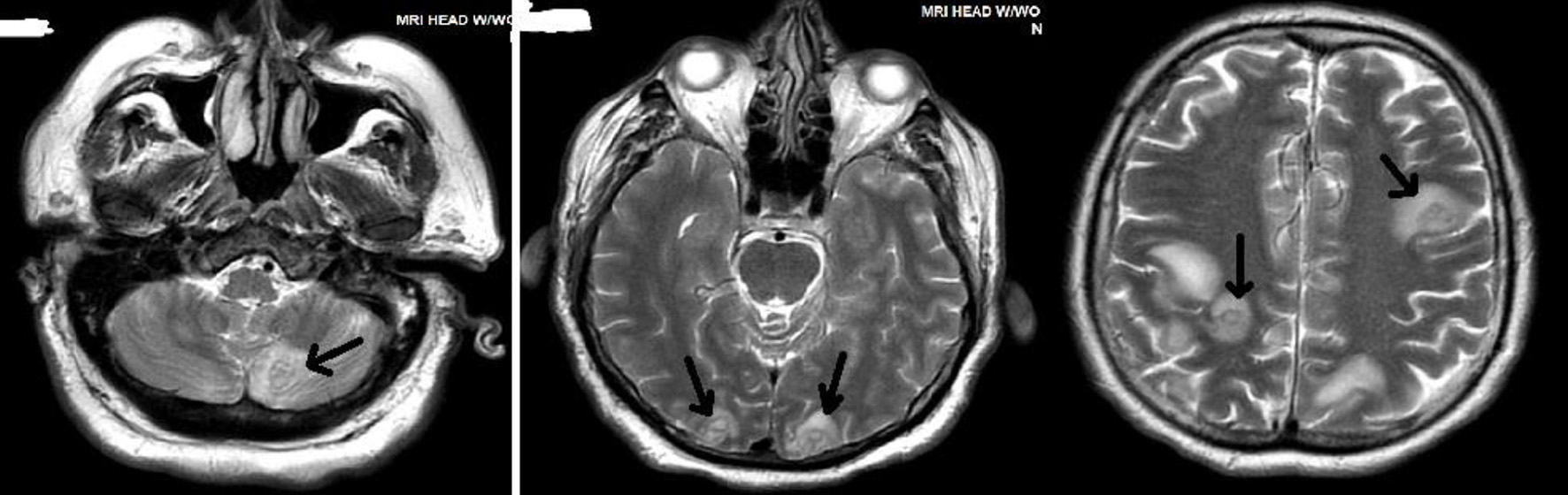
Figure 5. Magnetic resonance imaging of head with gadolinium showing central nervous system involvement of Nocardia. Black arrows show multiple ring-enhancing lesions in the cerebral and cerebellar hemispheres bilaterally.
Table
Table 1. Laboratory Values on Day 1 of Initial Hospital Admission
| Parameter | Value | Reference value |
|---|
| WBC: white blood cell count; CRP: C-reactive protein; pro-BNP: pro-b-type natriuretic peptide. |
| WBC (103/µL) | 15.3 | 4.0 - 11.2 |
| Erythrocyte sedimentation rate (mm/h) | 61 | 0 - 15 |
| Ferritin (ng/mL) | 2044.2 | 22 - 322 |
| High-sensitivity CRP (mg/dL) | > 10 | < 1.0 |
| Troponin (ng/mL) | 0.091 | 0.000 - 0.045 |
| D-dimer (µg/mL FEU) | 4.08 | 0 - 0.52 |
| Pro-BNP (pg/mL) | 909.0 | 0 - 125 |
| Hemoglobin A1c (%) | 13.7 | < 5.7% |