Figures
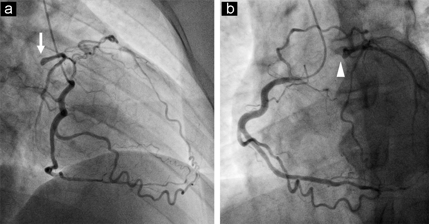
Figure 1. The left anterior oblique 45° view on coronary angiography shows the rich collateral flow from the right coronary artery to the left anterior descending coronary artery and circumflex artery with the stump of the LMCA (a, arrow), as does the right anterior oblique 35° view (b, arrowhead).
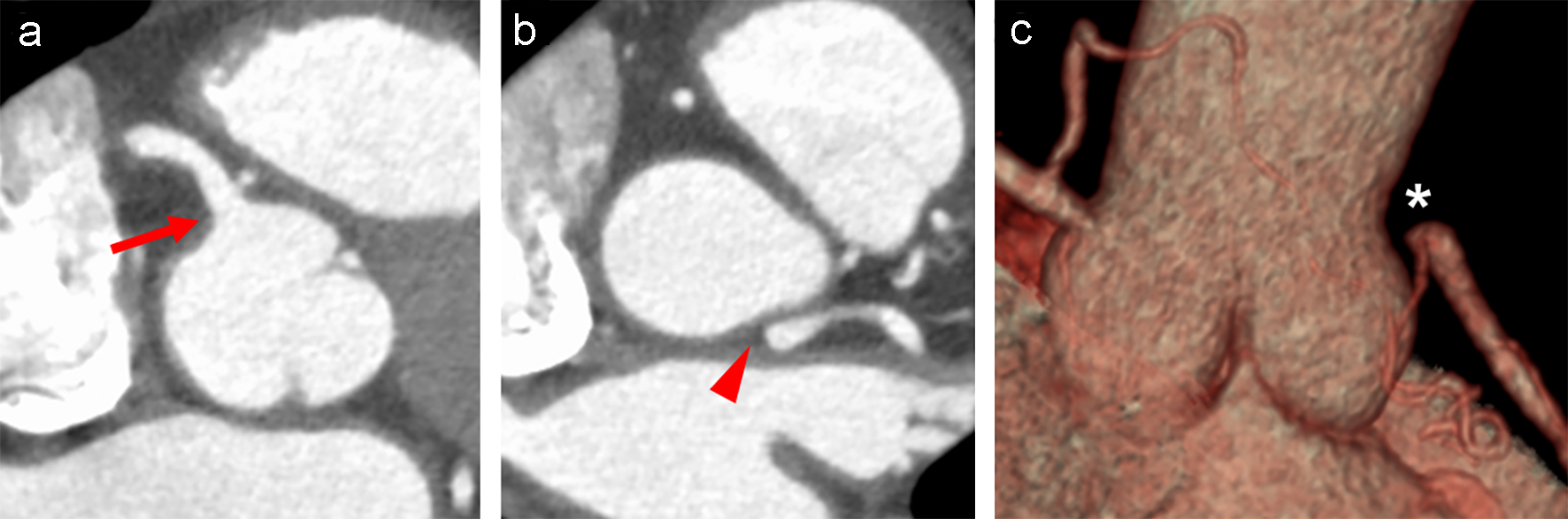
Figure 2. Short axial images of CT obtained after the administration of contrast material show a clear ostium of the right coronary artery (a, arrow), but no connection between the LMCA and the left sinus of Valsalva (b, arrowhead). Note that no trace indicative of the LMCA ostium is detected in the sinus of Valsalva. The absence of the LMCA is shown on the volume rendering image (c, asterisk). CT: computed tomography; LMCA: left main coronary artery.
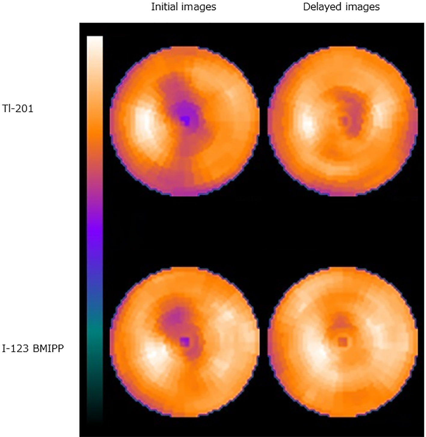
Figure 3. On dual isotope scintigraphy, the Tl-201 bull’s-eye map shows reduced tracer uptake in the anterior region to the apex on the initial image with fill-in on the delayed image or 3 h after injection, findings consistent with ischemic but viable myocardium in the region. Similarly, on the I-123 BMIPP bull’s-eye map, decreased fatty acid metabolism (i.e., myocardial ischemia) is noted in the same area. Note that reduced tracer uptake on the initial images is milder with I-123 BMIPP than with Tl-201.
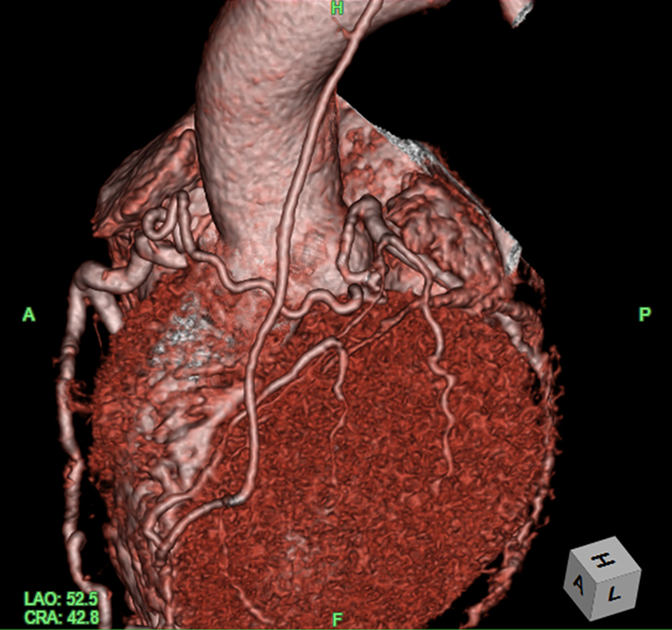
Figure 4. CT angiography after surgery shows that the left anterior descending coronary artery without the LMCA ostium is perfused by the bypassed graft and rich collateral flow from the right coronary artery. Image courtesy of Dr. Satsuki Fukushima. CT: computed tomography; LMCA: left main coronary artery.