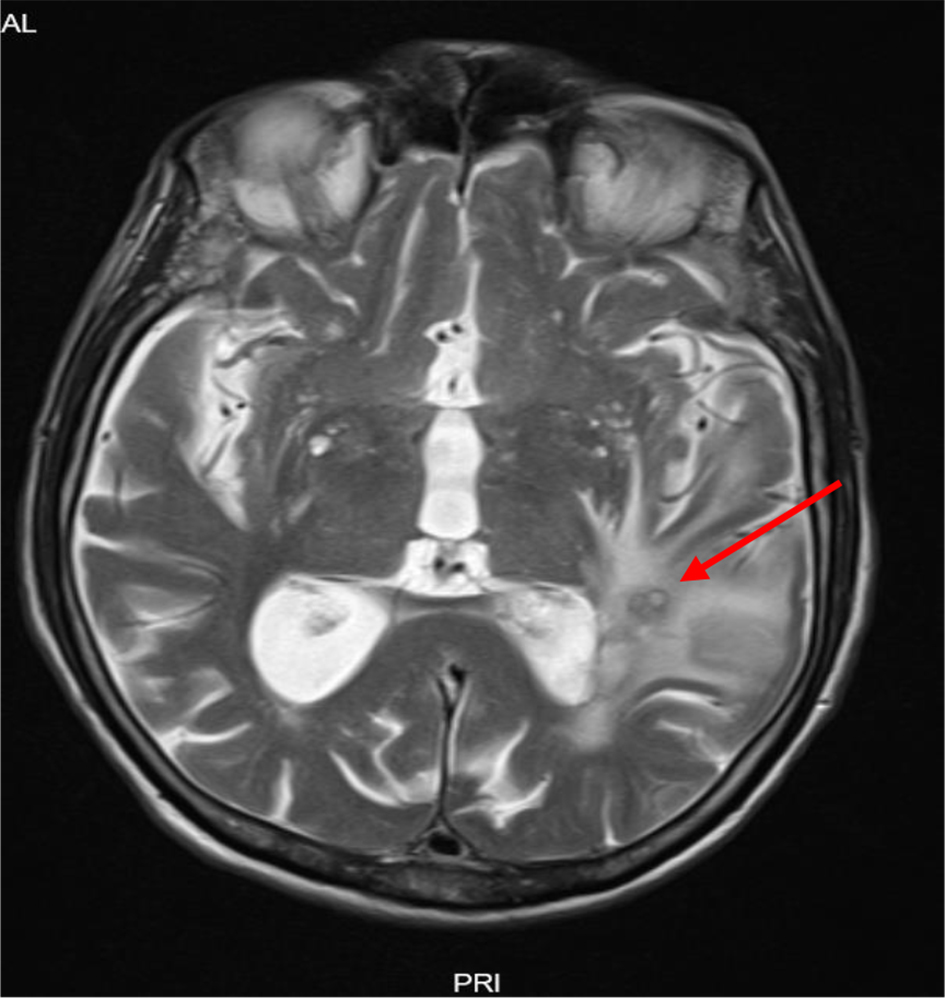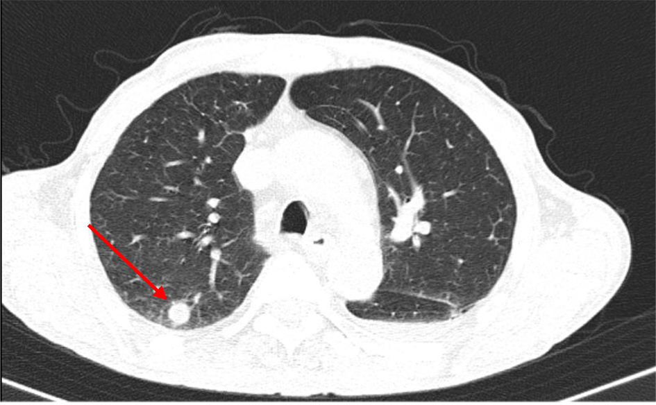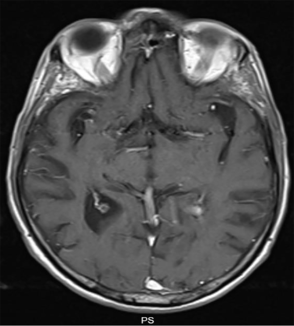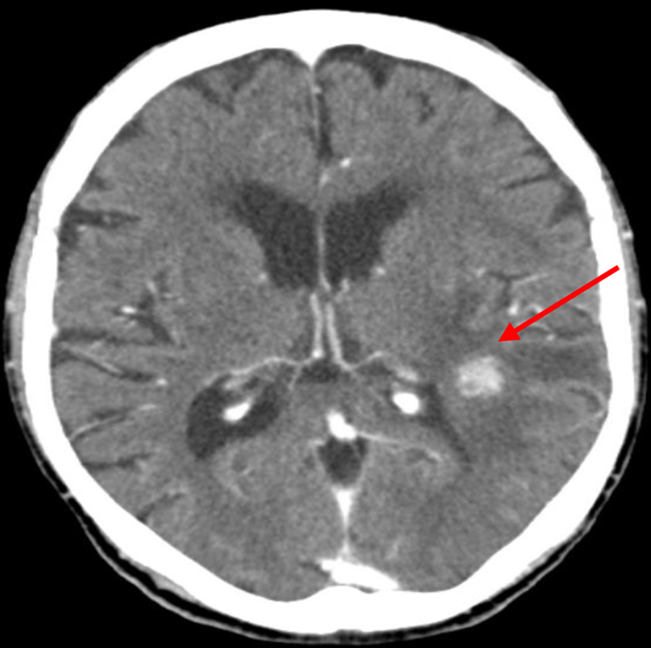
Figure 1. Admission cerebral computed tomography (CT) scan, showing a nodular lesion and surrounding edema (arrow).
| Journal of Medical Cases, ISSN 1923-4155 print, 1923-4163 online, Open Access |
| Article copyright, the authors; Journal compilation copyright, J Med Cases and Elmer Press Inc |
| Journal website https://www.journalmc.org |
Case Report
Volume 12, Number 5, May 2021, pages 205-208
Disseminated Nocardiosis: The Complexity of the Diagnosis
Figures