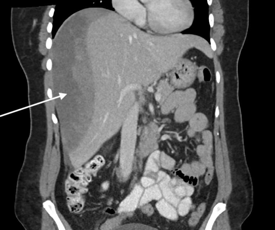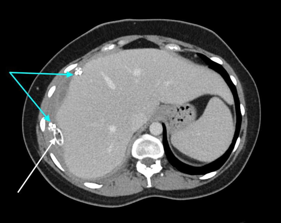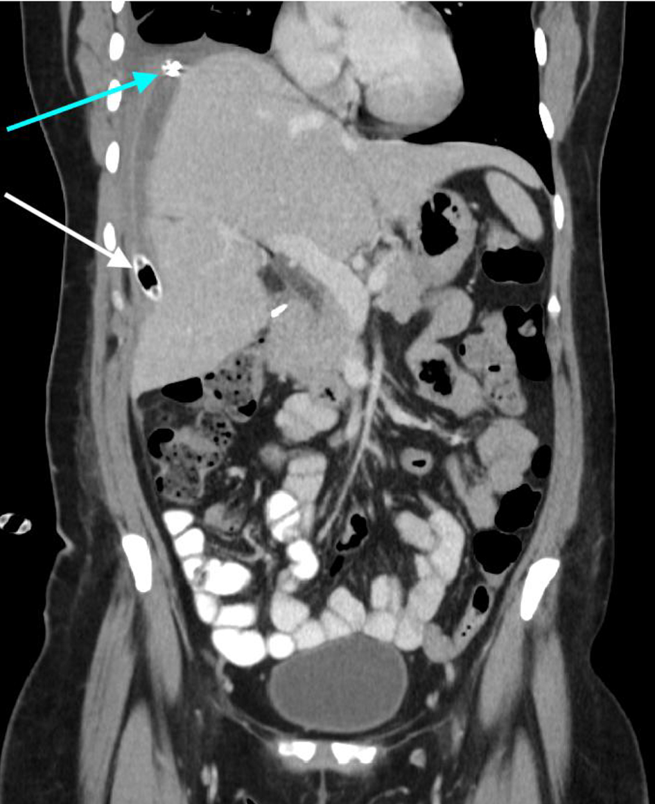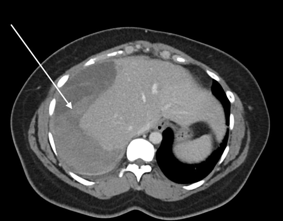
Figure 1. Axial CT demonstrating the large subcapsular hepatic hematoma involving most of the right lobe of the liver, highlighted by the arrow. CT: computed tomography.
| Journal of Medical Cases, ISSN 1923-4155 print, 1923-4163 online, Open Access |
| Article copyright, the authors; Journal compilation copyright, J Med Cases and Elmer Press Inc |
| Journal website https://www.journalmc.org |
Case Report
Volume 12, Number 5, May 2021, pages 186-189
Subcapsular Hepatic Hematoma Post-Endoscopic Retrograde Cholangiopancreatography Requiring Surgical Necrosectomy
Figures