Journal of Medical Cases, ISSN 1923-4155 print, 1923-4163 online, Open Access
Article copyright, the authors; Journal compilation copyright, J Med Cases and Elmer Press Inc
Journal website https://www.journalmc.org

Case Report

Volume 12, Number 5, May 2021, pages 181-185


Primary Breast Diffuse Large B-Cell Lymphoma in a 42-Year-Old Female: A Case Report and Review of Literature

Figures

Figure 1.
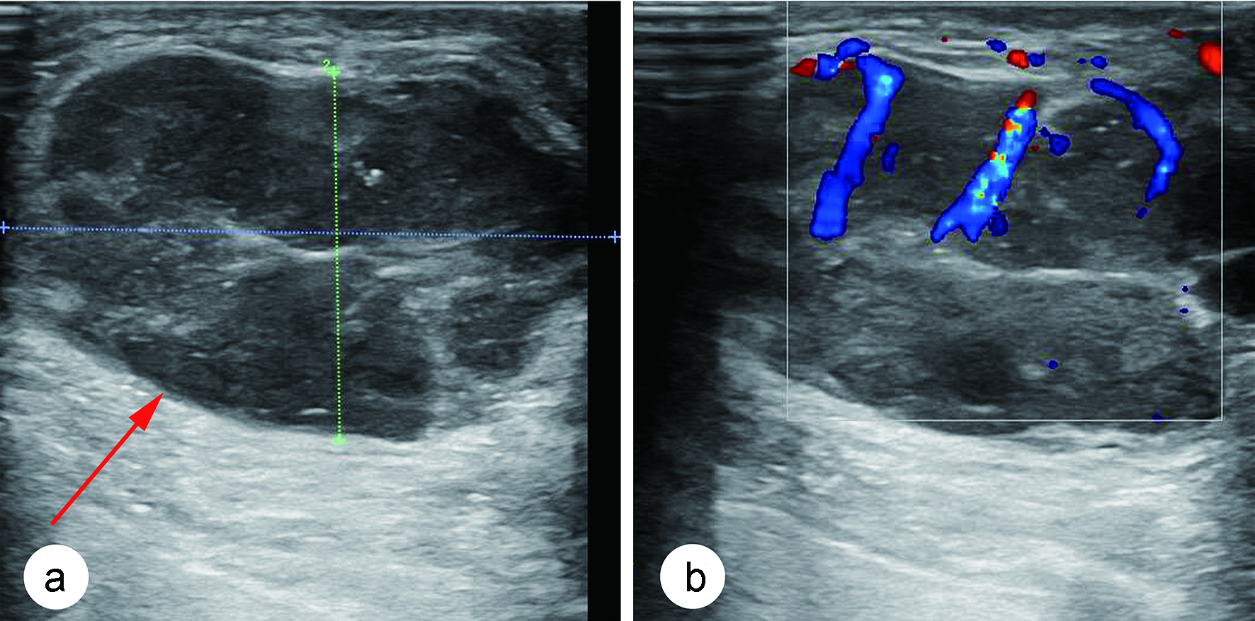
Figure 1. (a) Ultrasonography of breast showed an irregular hypoechoic nodule with multiple punctate calcifications in the left areola region (red arrow). (b) Color Doppler flow imaging revealed blood flow signals in this nodule and a high-impedance arterial flow pattern.
Figure 2.
Figure 2. Mammography of left breast showed a high-density mass with partially obscured margins (red arrows).
Figure 3.
Figure 3. Frozen pathological sections revealed that the tumor cells were morphologically diverse with abundant mitotic figures (hematoxylin & eosin (H&E), × 100).
Figure 4.
Figure 4. (a) Positron emission tomography-computed tomography (PET-CT) revealed an abnormally hypermetabolic mass of left breast (red arrows). (b) The hypermetabolic mass disappeared after four cycles of chemotherapy.