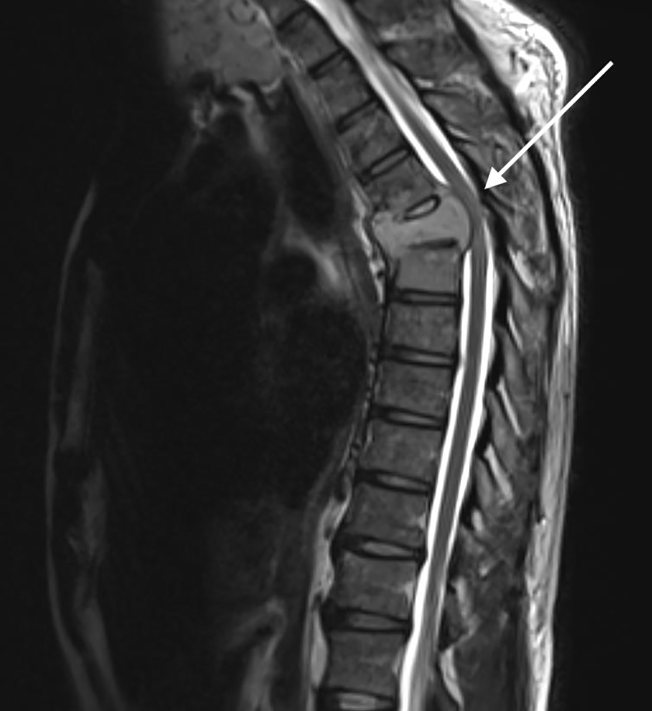
Figure 1. Sagittal magnetic resonance imaging (MRI) of the thoracic spine demonstrating a severe attenuation of the T5 thoracic cord with increased abnormal T2 signal (highlighted by the arrow).
| Journal of Medical Cases, ISSN 1923-4155 print, 1923-4163 online, Open Access |
| Article copyright, the authors; Journal compilation copyright, J Med Cases and Elmer Press Inc |
| Journal website https://www.journalmc.org |
Case Report
Volume 12, Number 5, May 2021, pages 177-180
Metastatic Follicular Thyroid Carcinoma Presenting as Thoracic Cord Compression
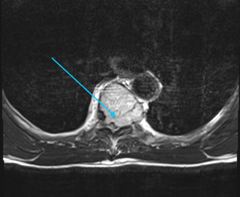
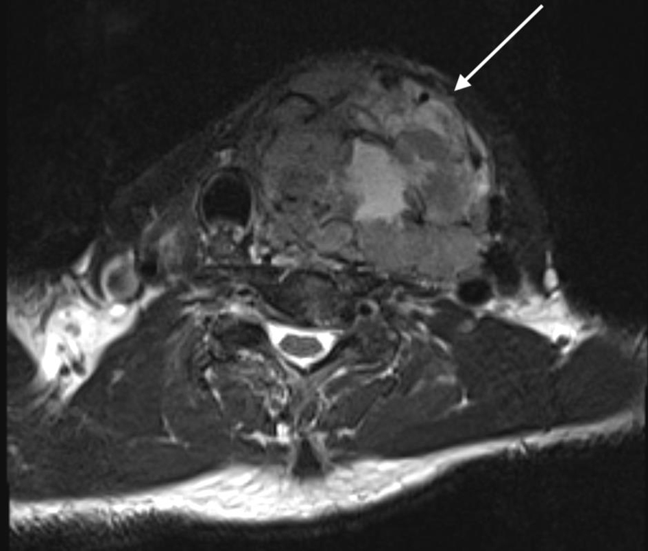
Figures